• Skip to primary navigation
  • Skip to main content
  • Skip to primary sidebar
  • Skip to footer

iBiology

Bringing the World's Best Biology to You

  • Start Here
    • Explore iBiology
    • All Playlists
    • All Speakers
    • All Talks
    • What’s new at iBiology?
  • Research Talks
    • Talks by Topic
      • Biochemistry
      • Bioengineering
      • Biophysics
      • Cell Biology
      • Development and Stem Cells
      • Ecology
      • Evolution
      • Genetics and Gene Regulation
      • Human Disease
      • Immunology
      • Microbiology
      • Neuroscience
      • Plant Biology
      • Techniques
      • Archive
    • Talks by Series
      • Bench to Bedside
      • Famous Discoveries
      • Great Unanswered Questions
      • Microscopy Series
      • Share Your Research Series
  • Stories
    • Background to Breakthrough
    • Interviews and Profiles
    • Science and Society
  • Courses
  • Careers
    • Professional Development Talks
    • Professional Development Courses
    • Career Exploration
    • NRMN Resources
    • Biomedical Workforce
  • Educators
  • About
    • Mission
    • Commitment to Inclusion
    • iBiology Team
    • Board of Directors
    • iBiology Funders and Partnerships
    • Contact Us
  • Donate
Home » Courses » Microscopy Series » Contrast Generation for Transmitted Light

Examples of Using Polarization Microscopy

  • Duration: 5:50
  • Downloads
    • Hi-Res
    • Low-Res
  • Subtitles
    • English
  • Transcript

00:00:12.06 Now, we can improve the fluorescent microscope even further, as you see here.
00:00:18.12 And with these, for example, ordinary polarization microscopes, we can have only a limited
00:00:26.14 image resolution because light... as it bends inside lenses, they get twisted,
00:00:34.07 and that messes up the polarized light.
00:00:36.10 So, what we did was to introduce what is known as a polarization rectifier, as is shown on
00:00:43.07 the right-hand side.
00:00:44.24 And the rectifier corrects for all this aberration and brings a completely dark field, even with
00:00:53.00 a high numerical aperture objective and condenser.
00:00:56.14 So, that means we can get much better sensitivity for detecting the weak birefringence.
00:01:02.05 Also, by using video, one can improve the contrast of polarized light and other kinds
00:01:08.20 of microscopes.
00:01:10.05 So, we again gain a great deal of detectability.
00:01:14.03 For example, here we see, in a time-lapse movie, the development of a...
00:01:18.26 a tunicate egg.
00:01:35.28 On the right side, we just saw the second polar body forming, and then within the...
00:01:42.13 near where the polar body formed is the female pronucleus migrating towards the male pronucleus.
00:01:48.28 Then they fuse and form the first division spindle, which is birefringent.
00:01:54.04 And then, after the spindle separates you get the two daughter cells.
00:01:58.07 Again, you see the big asters, there.
00:02:01.11 Then they form the daughter nuclei.
00:02:04.17 The daughter nuclei again form two cells, as you see here.
00:02:13.04 This is now in DIC.
00:02:15.04 The daughter nuclei migrate to the middle and form the four daughter cells.
00:02:20.22 This keeps on going and going, so that if we look at the next day, the same tunicate embryo
00:02:26.08 is a little tadpole that is swimming around.
00:02:29.02 And this is all done non-destructively and following the molecular changes inside living cells.
00:02:37.26 Here, I illustrate why we use a compensator.
00:02:42.00 By using a compensator, as we see down below, on the left-hand side, the spindle birefringence
00:02:48.24 comes out very clearly.
00:02:51.03 The compensator improves the sensitivity of the fluorescent microscope for detecting
00:02:58.12 weak birefringence.
00:03:00.03 And also it tells us which way the molecules are lined up.
00:03:03.08 For example, up and down, the spindles are dark; horizontally, they're bright.
00:03:08.02 That is because the protein molecules are lined up along the length of the spindle.
00:03:13.25 And also it will measure how many molecules are lined up by the birefringence, which can
00:03:21.02 be compensated by adjusting the compensator.
00:03:26.00 This also tells us which part of the cell has become birefringent.
00:03:31.14 In this case, the spindle has moved toward what's known as the vegetal pole,
00:03:37.24 and then the cells divide in order to separate the chromosomes into two so that, as you see on
00:03:45.25 the right-hand side, the result of the left-hand spindle displacement is the formation of
00:03:53.03 four tiny cells and four large cells.
00:03:56.07 And this is the beginning of cell differentiation.
00:03:59.05 The offspring of the tiny cells form the spicules -- the bones of the sea urchin that I've just mentioned --
00:04:07.08 and also the gonads, and the large ones form the mesenchyme.
00:04:11.20 In the next slide, we briefly see what we can see by looking at sperm structure.
00:04:19.09 For example, here is cave cricket sperm.
00:04:24.10 As you see you in the left-hand column, the top of the sperm head, you see black and white
00:04:29.17 domains.
00:04:30.20 What those are are little regions in which DNA molecules are tilted to the left and right.
00:04:38.06 And actually they are crucial to how chromosomes are arranged, how DNA molecules are
00:04:44.07 arranged inside chromosomes.
00:04:45.16 And by using polarized ultraviolet light, we can even find out what... within each gyre
00:04:53.11 of the chromosome, how the DNA is lined up.
00:04:57.04 And the far right, top picture shows how we concluded the DNA molecules to be lined up
00:05:05.25 within each of the chromosomes, which are very, very much smaller than what we can
00:05:11.18 resolve with the light microscope.
00:05:13.00 But these kind of things you can all find out by using polarized light.
00:05:17.24 So, that's a quick explanation of how we can use polarized light in some biological examples.
00:05:24.27 Thank you.

This Talk
Speaker: Shinya Inoue
Audience:
  • Researcher
Recorded: July 2011
More Talks in Microscopy Series
  • Microscopy Edward Salmon
    Pragmatics of DIC and Video-Enhanced Contrast Microscopy
  • Phase, Polarization, and DIC Stephen Ross
    Phase, Polarization, and DIC Microscopy Lab
  • Introduction to Fluorescence Microscopy
All Talks in Microscopy Series
Share

Talk Overview

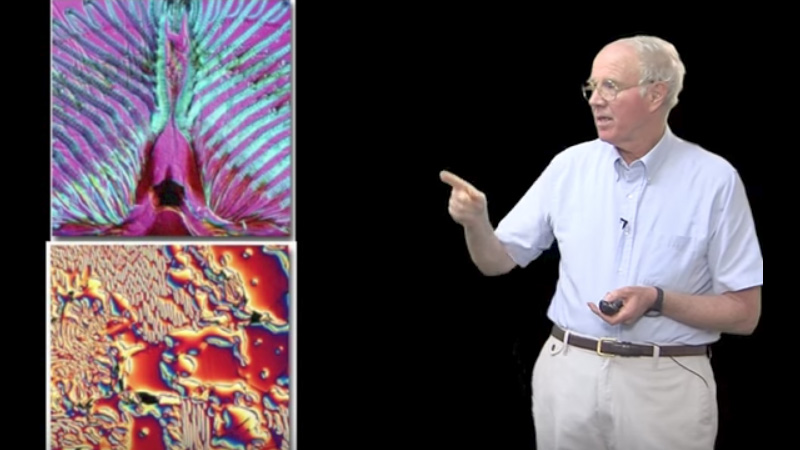
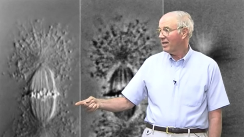
Shinya describes examples of using polarization microscopy to image living biological organisms, including watching processes of mitosis.

Speaker Bio

Shinya Inoue

Shinya Inoue

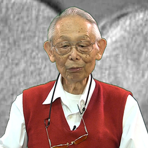
Dr. Inoue is a Distinguished Scientist at the Marine Biological Laboratory in Woods Hole, MA. He has made numerous contributions to microscopy and cell biology, including developing polarized light microscopy and video microscopy. Inoue was awarded the Order of the Sacred Treasure by the Government of Japan and is a member of the National Academy… Continue Reading

Playlist: Microscopy Series

  • Differential Interference Contrast (DIC) Microscopy Edward Salmon
    Differential Interference Contrast (DIC) Microscopy
  • Microscopy Edward Salmon
    Pragmatics of DIC and Video-Enhanced Contrast Microscopy
  • Phase, Polarization, and DIC Stephen Ross
    Phase, Polarization, and DIC Microscopy Lab
  • Introduction to Fluorescence Microscopy

Reader Interactions

Leave a Reply Cancel reply

Your email address will not be published. Required fields are marked *

Primary Sidebar

Sign up for the Science Communication Lab education newsletter

  • Sign up Newsletter
  • This field is for validation purposes and should be left unchanged.

Privacy Policy

Help us keep bringing the world’s best biology to you!

Footer

Funders

NSF
NIGMS
Lasker
Rita Allen

Start Here

  • Talks for Everyone
  • Talks for Students
  • Talks for Research
  • Talks for Educators

Explore

  • Explore
  • All Playlists
  • All Speakers
  • All Talks

Talks By Topic

  • Biochemistry
  • Bioengineering
  • Biophysics
  • Cell Biology
  • Development and Stem Cells
  • Ecology
  • Genetics and Gene Regulation
  • Human Disease
  • Immunology
  • Microbiology
  • Neuroscience
  • Plant Biology
  • Techniques

Talks by Series

  • Bench to Bedside
  • Famous Discoveries
  • Great Questions
  • Share Your Research Series

Career

  • Professional Development
  • Career Exploration
  • NRMN Resources
  • Biomedical Workforce

Courses

  • Microscopy Series
  • Short Microscopy Series
  • Open edX Courses
  • Cell Biology Flipped Course
  • Engineering Life Flipped Course
  • Evolution Flipped Course

Educator

  • Educator Registration
  • Educator Resources
  • Log In

About Us

  • About Us
  • iBiology Team
  • Wonder Collaborative
  • Contact Us
  • Mission
  • Privacy Policy
  • SCL Financial Conflict of Interest Policy

This material is based upon work supported by the National Science Foundation and the National Institute of General Medical Sciences under Grant No. 2122350 and 1 R25 GM139147. Any opinion, finding, conclusion, or recommendation expressed in these videos are solely those of the speakers and do not necessarily represent the views of the Science Communication Lab/iBiology, the National Science Foundation, the National Institutes of Health, or other Science Communication Lab funders.

© 2023 - 2006 iBiology · All content under CC BY-NC-ND 3.0 license · Privacy Policy · Terms of Use · Usage Policy
 

Power by iBiology