• Skip to primary navigation
  • Skip to main content
  • Skip to primary sidebar
  • Skip to footer

iBiology

Bringing the World's Best Biology to You

  • Start Here
    • Explore iBiology
    • All Playlists
    • All Speakers
    • All Talks
    • What’s new at iBiology?
  • Research Talks
    • Talks by Topic
      • Biochemistry
      • Bioengineering
      • Biophysics
      • Cell Biology
      • Development and Stem Cells
      • Ecology
      • Evolution
      • Genetics and Gene Regulation
      • Human Disease
      • Immunology
      • Microbiology
      • Neuroscience
      • Plant Biology
      • Techniques
      • Archive
    • Talks by Series
      • Bench to Bedside
      • Famous Discoveries
      • Great Unanswered Questions
      • Microscopy Series
      • Share Your Research Series
  • Stories
    • Background to Breakthrough
    • Interviews and Profiles
    • Science and Society
  • Courses
  • Careers
    • Professional Development Talks
    • Professional Development Courses
    • Career Exploration
    • NRMN Resources
    • Biomedical Workforce
  • Educators
  • About
    • Mission
    • Commitment to Inclusion
    • iBiology Team
    • Board of Directors
    • iBiology Funders and Partnerships
    • Contact Us
  • Donate
Home » Courses » Microscopy Series » Image Formation

Features of an Objective Lens

  • Duration: 4:40
  • Downloads
    • Hi-Res
    • Low-Res
  • Subtitles
    • English
  • Transcript

01:00:13.02 the nomenclature on an objective lens
01:00:15.25 and cover what some of the properties
01:00:17.27 are and what information is
01:00:19.14 printed on the front of that lens.
01:00:21.20 So, the objective lens,
01:00:23.24 as we've talked about before
01:00:25.05 is really the most important part
01:00:27.04 of the optical system.
01:00:28.08 Also one of the more expensive parts
01:00:30.12 of an optical system.
01:00:32.13 The information on the lens,
01:00:33.27 on the top you can see the manufacturer,
01:00:36.21 it'll tell you the aberration correction
01:00:39.13 for example, here it says "Plan Apo,"
01:00:41.29 and that stands for "Plan" flatfield correction,
01:00:46.16 and "Apo" standing for apochromatic correction,
01:00:50.02 so a very broad range of wavelengths
01:00:52.19 from the violet to the red.
01:00:53.24 Okay, the magnification,
01:00:56.18 which is shown here as 60x
01:00:58.25 is printed, as well as the color code,
01:01:02.06 in our case, blue, for 60x lenses.
01:01:05.24 So all 60x lenses are going to have
01:01:08.05 a blue band around them.
01:01:09.12 And then it shows you a slash, and a 0.95,
01:01:15.14 that's the numerical aperture of the lens.
01:01:17.22 The light gathering capability
01:01:20.13 and the resolution capability of that lens.
01:01:23.21 Then below that,
01:01:25.17 you have specialized optical properties,
01:01:27.22 in this case, it says "DIC M,"
01:01:30.00 that means that this lens is
01:01:32.05 of sufficient quality and low strain
01:01:34.20 that it can be used for
01:01:35.18 the technique of differential interference contrast.
01:01:37.26 Similarly, if you had a lens
01:01:40.26 that was good for phase contrast,
01:01:43.03 and had a phase ring in it,
01:01:44.14 it would tell you that,
01:01:45.13 it would have a "Ph" for phase on it.
01:01:48.18 "M" designates that this is a medium
01:01:52.15 numerical aperture lens,
01:01:55.18 different manufacturers have different nomenclature
01:01:58.05 for their types of DIC.
01:02:00.20 And that basically tells you which prism
01:02:02.26 in the microscope to match with that lens
01:02:05.08 to get differential interference contrast to work.
01:02:08.02 Below that, will show you the tube length,
01:02:11.15 and most, as you can see that right here.
01:02:14.14 The tube length focal length,
01:02:16.01 now most microscopes nowadays
01:02:18.20 are infinity corrected, and that's why
01:02:20.21 you see the infinity symbol there on that bottom line.
01:02:23.10 /0.11-0.25, which is the cover glass
01:02:30.05 thickness that that lens can work with.
01:02:33.05 In older microscopes, finite microscopes,
01:02:36.28 it would give you a focal length
01:02:38.24 to the tube lens,
01:02:39.22 such as, say 160 or 200 / 0.11.
01:02:46.02 So the working distance is then shown here
01:02:50.28 as "WD," and the working distance is
01:02:55.01 the actual free working distance
01:02:58.18 not including the coverslip
01:03:00.11 that you can focus into the specimen.
01:03:02.12 So working distance in the case here,
01:03:05.20 .13, shown here in millimeters,
01:03:10.17 a 130 microns that you can then focus
01:03:13.27 beyond the coverslip into the specimen.
01:03:16.03 So the important point there is to
01:03:18.02 know that that already is taking into account
01:03:20.25 the coverslip and that's how far
01:03:22.20 you can focus into the specimen
01:03:24.27 on the other side of your cover glass.
01:03:26.22 The cover glass thickness range
01:03:30.21 is important and you need to adjust
01:03:34.08 the correction collar to correct for
01:03:36.04 spherical aberration, due to the
01:03:38.28 variability in the cover glass thickness.
01:03:41.09 And then you have sort of this cone here
01:03:44.22 with the front element in it.
01:03:46.29 Often times if it's an oil immersion lens
01:03:49.15 or an immersion lens
01:03:50.07 with short working distance,
01:03:51.14 that will retract with a spring.
01:03:53.17 And the mounting thread here, generally in brass.
01:03:58.14 And I think those are pretty much
01:04:00.19 the key points that you can get from
01:04:02.23 reading the front of an objective lens.
01:04:05.13 There are many many different categories
01:04:07.02 of lenses, and every manufacturer though,
01:04:11.04 will have this sort of standard information.
01:04:14.08 Thank you.

This Talk
Speaker: Steve Ross
Audience:
  • Researcher
Recorded: March 2012
More Talks in Microscopy Series
  • How To Clean an Objective Lens (Kurt Thorn)
    How to Clean Objective Lenses and Filters
  • Stephen Ross
    Cleaning a Microscope: How to Find Dirt in Your Optical System
  • Light Sources Nico Stuurman
    Light Sources for Microscopy
All Talks in Microscopy Series
Share

Talk Overview

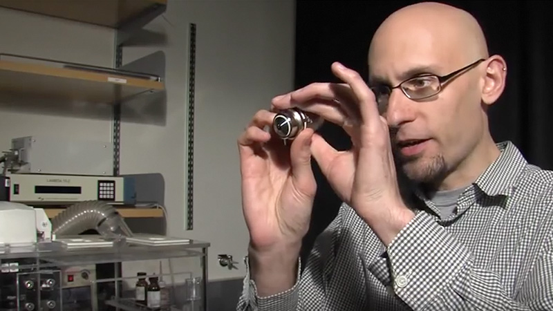
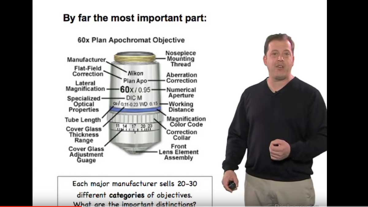
Steve Ross explains the features of an objective lens and what all of those markings actually mean.

Speaker Bio

Steve Ross

Steve Ross

Stephen Ross is the General Manager of Product and Marketing at Nikon Instruments.  He is also very involved in teaching microscopy at the Marine Biological Laboratory in Woods Hole and at the Bangalore Microscopy Course at the National Centre for Biological Sciences. Continue Reading

Playlist: Microscopy Series

  • Stephen Ross
    Eyepieces
  • How To Clean an Objective Lens (Kurt Thorn)
    How to Clean Objective Lenses and Filters
  • Stephen Ross
    Cleaning a Microscope: How to Find Dirt in Your Optical System
  • Light Sources Nico Stuurman
    Light Sources for Microscopy

Reader Interactions

Leave a Reply Cancel reply

Your email address will not be published. Required fields are marked *

Primary Sidebar

Sign up for the Science Communication Lab education newsletter

  • Sign up Newsletter
  • This field is for validation purposes and should be left unchanged.

Privacy Policy

Help us keep bringing the world’s best biology to you!

Footer

Funders

NSF
NIGMS
Lasker
Rita Allen

Start Here

  • Talks for Everyone
  • Talks for Students
  • Talks for Research
  • Talks for Educators

Explore

  • Explore
  • All Playlists
  • All Speakers
  • All Talks

Talks By Topic

  • Biochemistry
  • Bioengineering
  • Biophysics
  • Cell Biology
  • Development and Stem Cells
  • Ecology
  • Genetics and Gene Regulation
  • Human Disease
  • Immunology
  • Microbiology
  • Neuroscience
  • Plant Biology
  • Techniques

Talks by Series

  • Bench to Bedside
  • Famous Discoveries
  • Great Questions
  • Share Your Research Series

Career

  • Professional Development
  • Career Exploration
  • NRMN Resources
  • Biomedical Workforce

Courses

  • Microscopy Series
  • Short Microscopy Series
  • Open edX Courses
  • Cell Biology Flipped Course
  • Engineering Life Flipped Course
  • Evolution Flipped Course

Educator

  • Educator Registration
  • Educator Resources
  • Log In

About Us

  • About Us
  • iBiology Team
  • Wonder Collaborative
  • Contact Us
  • Mission
  • Privacy Policy
  • SCL Financial Conflict of Interest Policy

This material is based upon work supported by the National Science Foundation and the National Institute of General Medical Sciences under Grant No. 2122350 and 1 R25 GM139147. Any opinion, finding, conclusion, or recommendation expressed in these videos are solely those of the speakers and do not necessarily represent the views of the Science Communication Lab/iBiology, the National Science Foundation, the National Institutes of Health, or other Science Communication Lab funders.

© 2023 - 2006 iBiology · All content under CC BY-NC-ND 3.0 license · Privacy Policy · Terms of Use · Usage Policy
 

Power by iBiology