• Skip to primary navigation
  • Skip to main content
  • Skip to primary sidebar
  • Skip to footer

iBiology

Bringing the World's Best Biology to You

  • Start Here
    • Explore iBiology
    • All Playlists
    • All Speakers
    • All Talks
    • What’s new at iBiology?
  • Research Talks
    • Talks by Topic
      • Biochemistry
      • Bioengineering
      • Biophysics
      • Cell Biology
      • Development and Stem Cells
      • Ecology
      • Evolution
      • Genetics and Gene Regulation
      • Human Disease
      • Immunology
      • Microbiology
      • Neuroscience
      • Plant Biology
      • Techniques
      • Archive
    • Talks by Series
      • Bench to Bedside
      • Famous Discoveries
      • Great Unanswered Questions
      • Microscopy Series
      • Share Your Research Series
  • Stories
    • Background to Breakthrough
    • Interviews and Profiles
    • Science and Society
  • Courses
  • Careers
    • Professional Development Talks
    • Professional Development Courses
    • Career Exploration
    • NRMN Resources
    • Biomedical Workforce
  • Educators
  • About
    • Mission
    • Commitment to Inclusion
    • iBiology Team
    • Board of Directors
    • iBiology Funders and Partnerships
    • Contact Us
  • Donate
Home » » Advances in Optics and Nanoparticle Technology: Pushing the Boundaries of Light Microscopy

Advances in Optics and Nanoparticle Technology: Pushing the Boundaries of Light Microscopy

  • Duration: 16:59
  • Downloads
    • Hi-Res
    • Low-Res
  • Subtitles
    • English
  • Transcript

00:00:08.11 00:00:10.14 Hi I'm Steve Chu.
00:00:10.15 00:00:14.06 I'm a Professor at Stanford and I'm here to talk to you about some great questions in
00:00:14.07 00:00:21.07 life sciences at the intersection of biology physics and computation.
00:00:21.08 00:00:26.29 So let me start by posing a general question how do cells talk to one another for example
00:00:27.00 00:00:34.18 how do neurons in the brain talk to one another but at an organism level in real-time how
00:00:34.19 00:00:39.26 do cells signal each other to know when to divide and proliferate as to be sociable and
00:00:39.27 00:00:44.26 just sit tight and stay where you are when they divide and proliferate without outside
00:00:44.27 00:00:47.17 signals that's the cause of cancer.
00:00:47.18 00:00:51.10 And in answering these questions there's a further question what new measurement capabilities
00:00:51.11 00:00:58.18 do we have that we didn't previously have that would enable us to actually gain some
00:00:58.19 00:01:01.16 new insight into these questions.
00:01:01.17 00:01:06.22 So let me begin by quoting the Great American philospher of the 20th century in case you
00:01:06.23 00:01:12.03 don't know who that was it was Yogi Berra or is and he said many wise things
00:01:12.04 00:01:19.19 For example if you come to a fork in the road take it but I'm going to be referring to as
00:01:19.20 00:01:24.29 he said you can observe a lot by just watching and indeed imaging in biology and biomedicine
00:01:25.00 00:01:33.14 have had profound impacts on these fields and if you look at this list x-ray Imaging
00:01:33.15 00:01:40.04 electron microscopy NMR imaging all these things were honored by Nobel Prize because
00:01:40.05 00:01:45.05 they really revolutionize medicine and biology and discoveries made with these new instruments.
00:01:45.06 00:01:53.05 And in fact even last year the lowly venerable Optical microscope was again recognized by
00:01:53.06 00:01:59.25 its ability to look at single fluorescent molecules and also to do what we call sub-wavelength
00:01:59.26 00:02:01.15 optical resolution.
00:02:01.16 00:02:07.28 What that means is if you have a single point meter, let's say a fluorescent dye.
00:02:07.29 00:02:12.06 It usually is resolved into a blob about 250 nanometers in diameter.
00:02:12.07 00:02:18.06 It's the blur circle that's dictated by optical resolution, but if you ask a different question
00:02:18.07 00:02:22.14 where is the center of each of these blobs if you can see them one at a time you can
00:02:22.15 00:02:28.10 find the center to much better precision and it was that discovery that led W.E.
00:02:28.11 00:02:34.05 Moerner andEric Betzig to their portion of the Nobel Prize a similar type of related
00:02:34.06 00:02:39.07 super resolution imaging was also awarded to Stefan Hell.
00:02:39.08 00:02:42.26 Let me show you how much better it is.
00:02:42.27 00:02:48.27 On the left hand side in the first seminal paper Eric Betzig shows an image of fluorescence
00:02:48.28 00:02:50.28 from a cell.
00:02:50.29 00:02:56.05 This is your typical optical image the best optical image you can get And with this new
00:02:56.06 00:03:01.18 super resolution using on the right hand side what you're capable of doing In fact if you
00:03:01.19 00:03:07.13 zoom in on that right hand picture what you find is that the resolution is more than 10
00:03:07.14 00:03:13.18 times better than the optical image In fact you can visualize the location of about 51,000
00:03:13.19 00:03:22.25 individual molecules Here is another contribution that Eric Betzig made It had to do with a
00:03:22.26 00:03:28.00 different form of illumination slightly better than the normal optical resolution but enabling
00:03:28.01 00:03:34.09 Eric and his collaborators to take data so quickly that you can actually see in this
00:03:34.10 00:03:41.18 picture an embryo developing as you look in time and so this is now a three-dimensional
00:03:41.19 00:03:46.28 image of an embryo moving in time.
00:03:46.29 00:03:50.29 So let me step back and tell you what this is about Suppose you have a car that's analogous
00:03:51.00 00:03:56.28 to a single cell Now you may know what all the parts are and traditional biochemistry
00:03:56.29 00:03:59.29 molecular biology has given us a great parts list but in addition to that you may even
00:04:00.00 00:04:06.17 know that some of the parts for example the circle here the engine parts are part of the
00:04:06.18 00:04:12.05 engine but that doesn't really tell you how this cell works.
00:04:12.06 00:04:18.04 What you really want to know is what is the location of those molecules in conjunction
00:04:18.05 00:04:22.02 with each other and that will give you better insight into how the cell works so imagine
00:04:22.03 00:04:28.15 the pistons and valves and the cams and everything else as individual molecules Can you see if
00:04:28.16 00:04:36.10 molecular resolution in real-time this thing happening and by real-time I mean at the time
00:04:36.11 00:04:42.13 scales relevant to biology Can you actually see the parts moving We are unable to do this
00:04:42.14 00:04:47.03 today with conventional optical microscopy and conventional probes
00:04:47.04 00:04:56.06 So here's my dream If you have a method of Imaging for example magnetic resonance imaging
00:04:56.07 00:05:00.26 and here you have Homer Simpson's brain be imaged, there's also another form of imaging
00:05:00.27 00:05:06.13 where you can have Homer Simpson when he's thinking about certain things and as he thinks
00:05:06.14 00:05:12.15 about these things certain portions of the brain light up in functional magnetic resonance
00:05:12.16 00:05:18.06 imaging for example if Homer thinks about doughnuts which occupy a large part of his
00:05:18.07 00:05:19.24 brain that portion of his brain lights up.
00:05:19.25 00:05:29.08 So again we have beginning clues of how the brain is going to work but this is crude location
00:05:29.09 00:05:34.24 We want to do better than that On the other hand there have been exquisite things that
00:05:34.25 00:05:41.02 have been invented for example in the early mid-1990's by Neher and Sakmann where they
00:05:41.03 00:05:46.20 took a micropipette and they put it onto a single neuron and when they do this they can
00:05:46.21 00:05:51.04 actually measure through measuring currents they go in and out of the neuron individual
00:05:51.05 00:05:57.13 voltage spikes These voltage spikes are used by a neuron to release neurotransmitters that
00:05:57.14 00:06:03.10 go from the axon of one neuron to across the synapse to the dendrite of another neuron.
00:06:03.11 00:06:10.10 This is fantastic It revolutionized neurobiology This is an example of the voltage spikes - individual
00:06:10.11 00:06:15.27 voltage spikes being transmitted down on a transmission line of the neuron It can resolve
00:06:15.28 00:06:22.03 in time what is actually happening but it can only do one neuron you're working hard
00:06:22.04 00:06:29.27 a dozen neurons maybe 15 neurons but that's not a brain by a long shot
00:06:29.28 00:06:35.08 In a brain for example neurons connected to muscles or on the right hand side these are
00:06:35.09 00:06:40.21 sets of neurons connected to other neurons we have lovely static images but we want better
00:06:40.22 00:06:47.13 We want the moving pistons the fluorescent probes turn out to be a limiting factor for
00:06:47.14 00:06:55.07 the dyes we have you only collect about 10,000 maybe a hundred thousand photons before the
00:06:55.08 00:07:01.04 dyes are destroyed It takes 10 to 100 seconds to form an image and the best resolution pictures
00:07:01.05 00:07:06.13 that I showed you were actually taken with cells that are fixed they're dead they're
00:07:06.14 00:07:12.18 not moving And so is it possible to develop nanoprobes
00:07:12.19 00:07:19.15 that give you that same spatial resolution or you can follow the organisms and proteins
00:07:19.16 00:07:26.10 indefinitely so these are stable probes that just last forever and not only that can the
00:07:26.11 00:07:31.24 probes be extremely bright that is to say they give up so many photons that in a brief
00:07:31.25 00:07:42.08 instant of time you can capture essentially in freeze frame some very fast dynamics And
00:07:42.09 00:07:47.03 the extreme version of this was pioneered by Doc Edgerton of MIT where he used very
00:07:47.04 00:07:54.06 bright strobe lights and he could actually take a picture of a bullet piercing a playing
00:07:54.07 00:07:57.13 card So it's all about speed in terms of freezing
00:07:57.14 00:08:06.02 the motion Now we are working on a new set of probes One of them are little diamonds
00:08:06.03 00:08:11.11 molecular size diamonds to nano-sized diamonds And we start with these little seeds that
00:08:11.12 00:08:16.14 remarkably are actually found as a byproduct of oil refinery And you take these little
00:08:16.15 00:08:23.06 blue and green little seeds you put them on the surface and you can actually deposit carbon
00:08:23.07 00:08:30.08 and it will form into a diamond And here on the left hand side you see a picture of a
00:08:30.09 00:08:37.20 large molecular diamond maybe 1 micron in diameter where you clearly see the diamond
00:08:37.21 00:08:45.08 facets but we can actually make diamonds as small as 5 nanometers 10 nanometers This is
00:08:45.09 00:08:52.02 diamonds now only a dozen atoms across and here on the right hand side you see a transmission
00:08:52.03 00:08:58.24 electron image and it's called the lattice image because you're actually imaging columns
00:08:58.25 00:09:04.05 of individual atoms and you see that it is the structure of a dime We'd additionally
00:09:04.06 00:09:10.13 put impurities in these diamonds - silicon impurities and it turns out when you do this
00:09:10.14 00:09:16.07 they can actually emit a lot of light This is one example of a single particle 8 nanometers
00:09:16.08 00:09:21.25 in diameter emitting 10 million photons a second Actually they're not emitting 10 million
00:09:21.26 00:09:27.17 photons a second We're counting 10 million photons a second So it's really like a 100
00:09:27.18 00:09:33.24 million So with this very very bright source also photostable we think we can really make
00:09:33.25 00:09:47.08 an impact possibly on the imaging efforts And right now we're working on the ability [00:09:39,54] to actually get these diamonds modify them in a way so that they can actually be voltage
00:09:47.09 00:09:50.04 sensitive If they can be voltage sensor you can imagine putting them in particular neurons
00:09:50.05 00:09:56.10 in the brain thousands of them in real-time motion pictures you can see the neurons talk
00:09:56.11 00:10:01.06 to one another Another type of nanoparticle we think has
00:10:01.07 00:10:03.05 promise are putting impurity atoms.
00:10:03.06 00:10:11.17 These are rare impurities - thullium neodymium ytterbium into crystal structures and they
00:10:11.18 00:10:18.04 luminesce at different colors Now there are two advantages of these particles they're
00:10:18.05 00:10:23.27 not going to be as bright as diamonds but they're multi-colored number one and they
00:10:23.28 00:10:28.01 actually give off light in very long times 40 to 100 microseconds Most of the fluorescence
00:10:28.02 00:10:33.22 that comes from a cell the background color light is instantaneous It comes out in a billionth
00:10:33.23 00:10:38.24 of a second and so I'll tell you why we're interested in these.
00:10:38.25 00:10:56.03 For example, you can take a particle and you can go with thullium. [00:10:41,33] It emits this color or you can pick another particle that emits a different impurity at [00:10:47,79] that color or you can pick a particle that only has one color or the other color or you
00:10:56.04 00:10:59.09 can take another particle that has a different ratio of colors or you can take another particle
00:10:59.10 00:11:06.03 with a different ratio of colors and you can build up combinatorially each class of particles
00:11:06.04 00:11:11.21 with different ratios different colors means you can target many different proteins.
00:11:11.22 00:11:15.19 If you count up how many you think we can target it turns out to be about 39 different
00:11:15.20 00:11:16.01 proteins.
00:11:16.02 00:11:25.19 So imagine fluorescent labels each color denoting the piston rod the crankshaft the camshaft
00:11:25.20 00:11:28.04 all these different parts So this is the dream we don't know if we'll
00:11:28.05 00:11:35.07 get there but it's what it's about Now there is another thing that has been good
00:11:35.08 00:11:41.08 in biology that was actually started in astronomy.
00:11:41.09 00:11:53.03 Here on the left-hand side picture you see a green laser that is actually shining up 00:11:46.55] into the upper atmosphere and there's a thin sodium layer in this upper atmosphere
00:11:53.04 00:11:57.19 And when this laser hits it it actually emits yellow light It's bright enough It's called
00:11:57.20 00:11:58.26 a guide star.
00:11:58.27 00:12:04.17 Now all the atmospheric turbulence actually jumbles up the light to make the lights twinkle
00:12:04.18 00:12:05.23 but you don't want the twinkle.
00:12:05.24 00:12:12.11 So they do an adjustment of this mirror so it actually compensates for the twinkle and
00:12:12.12 00:12:18.25 it shows up as a bride single point of light and they use that to correct for the atmospheric
00:12:18.26 00:12:19.11 turbulence.
00:12:19.12 00:12:24.18 In microscopy there's an analogous situation here on the right hand side you see light
00:12:24.19 00:12:32.06 entering into a perfectly good view scene it's like a piece of glass or some water and
00:12:32.07 00:12:38.16 the wavefronts are even and you see a single point determined by the optical resolution
00:12:38.17 00:12:45.13 But if light propagates through and it's scatteringing off of parts of the cells - cells themselves
00:12:45.14 00:12:51.20 - the nuclei of cells the wavefront gets distorted The question is can you make a guide star
00:12:51.21 00:12:57.18 and so did you have the star and said okay it's the equivalent of twinkling it's distorted
00:12:57.19 00:13:01.28 it looks bigger and then you reshape before the light actually goes in so you can correct
00:13:01.29 00:13:08.06 for that and here is a picture of some very recent work done by scientists at Howard Hughes
00:13:08.07 00:13:14.10 Medical Institute where they on the left hand side you see a picture when you don't have
00:13:14.11 00:13:21.06 this correction and it's a little bit blurry and on the right hand side you see a picture
00:13:21.07 00:13:25.18 where it's been corrected Now what you're actually looking at is another
00:13:25.19 00:13:32.20 advance These are optical probes that can sense changes in calcium in a cell so these
00:13:32.21 00:13:39.07 are neurons in a live brain and as you see this movie what you see on the right hand
00:13:39.08 00:13:45.06 side is much clearer definition of what's going on after you've taken out the scattering
00:13:45.07 00:13:49.04 light distortion Now this is great work They were able to look
00:13:49.05 00:13:55.05 500 microns deep into tissue or what was its limitation The signal was coming out of green
00:13:55.06 00:14:01.16 light and you can't look further than 500 microns It gets absorbed by the tissue
00:14:01.17 00:14:07.17 What's good about our rare earth is they are giving out light where it's an order of magnitude
00:14:07.18 00:14:15.05 less attenuated than the green light 10 times further and not only that it's giviing out
00:14:15.06 00:14:21.26 light that is completely devoid of background scattered light of light scattering of order
00:14:21.27 00:14:25.18 of fluorescence from the surrounding tissue because the light we're looking at comes out
00:14:25.19 00:14:32.29 in 100 microseconds not 1 nanosecond therefore you have a guide star that's nearly perfect
00:14:33.00 00:14:38.05 and with that you can make much deeper corrections and because you're using the right wavelength
00:14:38.06 00:14:43.13 in the most transparent part of tissue you can see much deeper
00:14:43.14 00:14:53.08 So we think perhaps you can look 3, 5, 6 millimeters into tissue and the question is can you use
00:14:53.09 00:15:00.03 adaptive optics looking a half a centimeter into tissue that gives you cellular resolution
00:15:00.04 00:15:04.29 and that would be really wonderful because if you can do that you can track the migration
00:15:05.00 00:15:09.26 of individual cells you can have cellular resolution deep into tissue that you simply
00:15:09.27 00:15:14.19 didn't have before It could be very useful for cancer stem cell research all these other
00:15:14.20 00:15:20.06 things So in summary if you look back the optical
00:15:20.07 00:15:26.12 microscope which is really developed in the 1600s Leeuwenhoek as you see was the first
00:15:26.13 00:15:30.24 Master at building really great lenses so that you can actually see things
00:15:30.25 00:15:38.07 There were other people but their microscopes weren't as good and someone discovered this,
00:15:38.08 00:15:44.10 encouraged Leeuwenhoek to submit a paper to the Royal Society which he did and this is
00:15:44.11 00:15:50.27 the final lesson He gets a reply back from his submission invited submission.
00:15:50.28 00:15:56.27 Dear Mr. van Leeuwenhoek, Your letter of October 10th has been received here with amusement.
00:15:56.28 00:16:01.04 Your account of myriad quote little animals seen swimming in rainwater led one member
00:16:01.05 00:16:08.24 to imagine that your might have contained an ample portion of distilled spirits imbibed
00:16:08.25 00:16:09.24 by the investigator.
00:16:09.25 00:16:12.03 It has been decided not to publish your communication.
00:16:12.04 00:16:18.17 However all here wish your little animals health and good husbandry by their ingenous
00:16:18.18 00:16:20.20 discoverer.
00:16:20.21 00:16:23.12 So that was a rejection.
00:16:23.13 00:16:30.03 So as you think about what you're doing, prepare to be rejected but stay the course and have
00:16:30.04 00:16:34.04 a vision for something that can transform science.
00:16:34.05 00:16:58.10 Thank you.

This Talk
Speaker: Steven Chu
Audience:
  • Researcher
Recorded: June 2015
More Talks in Microscopy Series
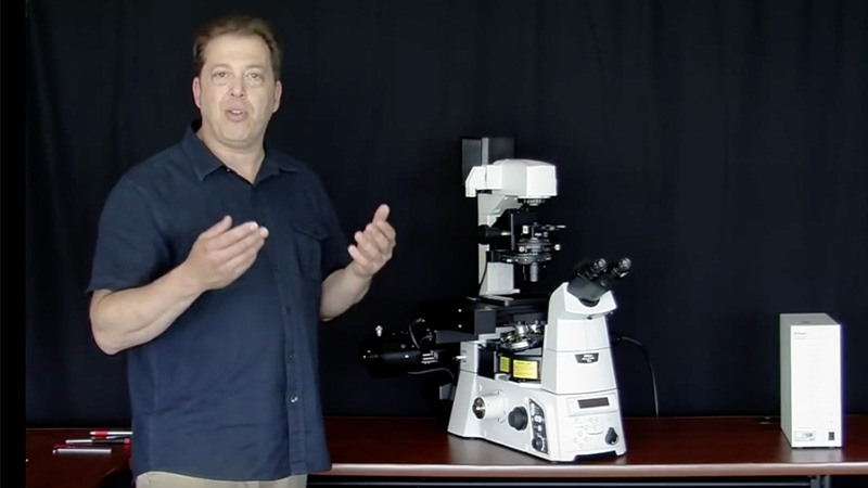
  • Disassembling a Nikon Ti Eclipse Stephen Ross
    Demystifying Microscopes: Disassembling a Nikon Ti Eclipse Microscope
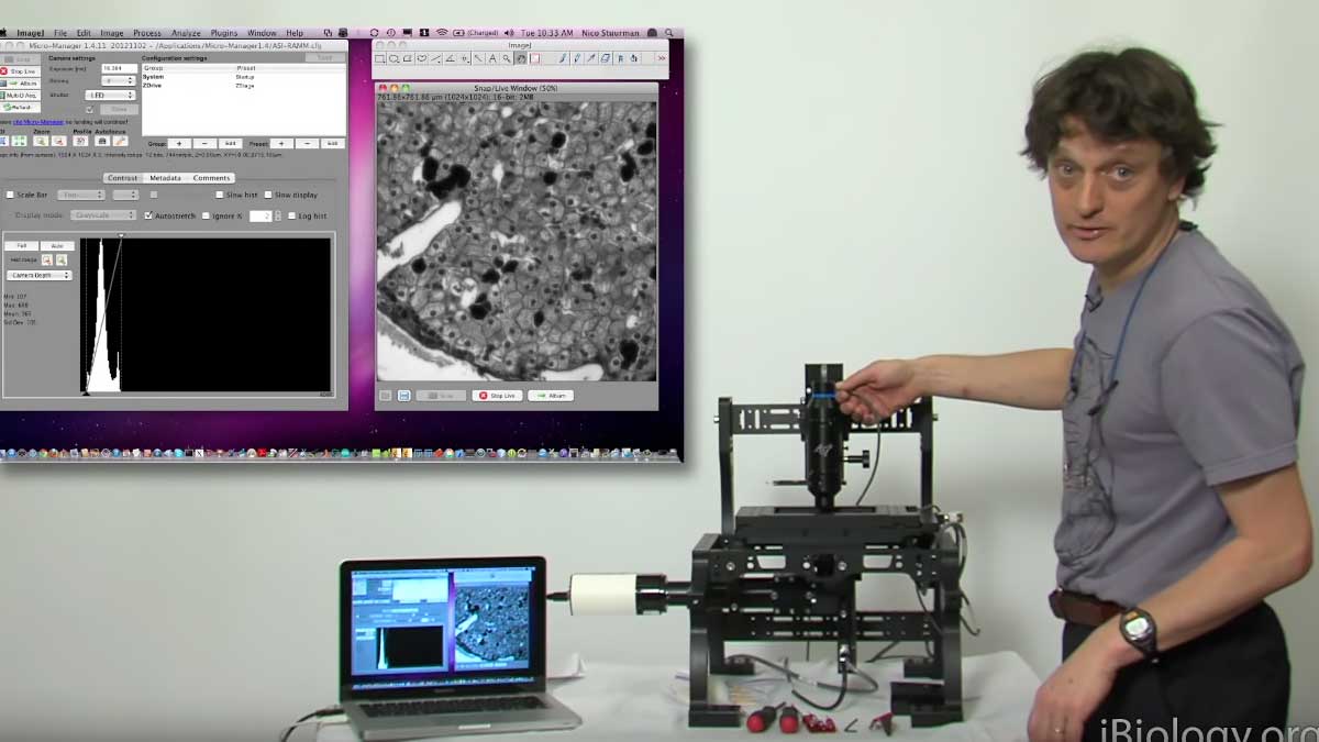
  • Nico Sturman on iBiology: Disassembling and ASI Ramm in Microscopy
    Demystifying Microscopes: Disassembling an ASI RAMM Microscope
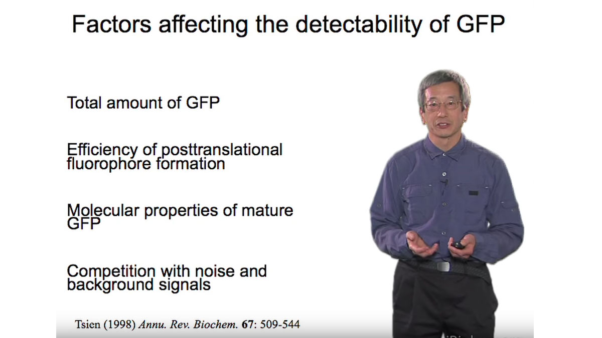
  • Optimizing Detection of GFP
All Talks in Microscopy Series
Share

Talk Overview

In order to understand how cells function at the molecular level in an organism, we need to observe the location and behavior of single molecules inside living tissue. Conventional optical microscopy and fluorescent probes/markers fall short in allowing us to do this. However, Steven Chu discusses recent advances in optics and nanoparticle technology, which hold great promise for observing individual proteins in living cells and tissues in real-time.

Speaker Bio

Steven Chu

Steven Chu

Steven Chu is Professor of Physics and Molecular & Cellular Physiology and the William R. Kenan, Jr., Professor of Humanities and Sciences at Stanford University. From 2009 to 2013, he served as US Secretary of Energy. At the time of his appointment to the Cabinet, he was a Professor of Physics and Molecular and Cell… Continue Reading
Playlist: Microscopy Series

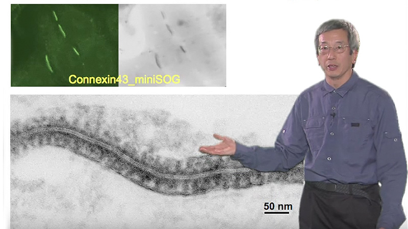
  • Correlating Fluorescence With Electron Microscopy Roger Tsien
    Correlating Fluorescence with Electron Microscopy
  • Disassembling a Nikon Ti Eclipse Stephen Ross
    Demystifying Microscopes: Disassembling a Nikon Ti Eclipse Microscope
  • Nico Sturman on iBiology: Disassembling and ASI Ramm in Microscopy
    Demystifying Microscopes: Disassembling an ASI RAMM Microscope
  • Optimizing Detection of GFP

Reader Interactions

Leave a Reply Cancel reply

Your email address will not be published. Required fields are marked *

Primary Sidebar

Sign up for the Science Communication Lab education newsletter

  • Sign up Newsletter
  • This field is for validation purposes and should be left unchanged.

Privacy Policy

Help us keep bringing the world’s best biology to you!

Footer

Funders

NSF
NIGMS
Lasker
Rita Allen

Start Here

  • Talks for Everyone
  • Talks for Students
  • Talks for Research
  • Talks for Educators

Explore

  • Explore
  • All Playlists
  • All Speakers
  • All Talks

Talks By Topic

  • Biochemistry
  • Bioengineering
  • Biophysics
  • Cell Biology
  • Development and Stem Cells
  • Ecology
  • Genetics and Gene Regulation
  • Human Disease
  • Immunology
  • Microbiology
  • Neuroscience
  • Plant Biology
  • Techniques

Talks by Series

  • Bench to Bedside
  • Famous Discoveries
  • Great Questions
  • Share Your Research Series

Career

  • Professional Development
  • Career Exploration
  • NRMN Resources
  • Biomedical Workforce

Courses

  • Microscopy Series
  • Short Microscopy Series
  • Open edX Courses
  • Cell Biology Flipped Course
  • Engineering Life Flipped Course
  • Evolution Flipped Course

Educator

  • Educator Registration
  • Educator Resources
  • Log In

About Us

  • About Us
  • iBiology Team
  • Wonder Collaborative
  • Contact Us
  • Mission
  • Privacy Policy
  • SCL Financial Conflict of Interest Policy

This material is based upon work supported by the National Science Foundation and the National Institute of General Medical Sciences under Grant No. 2122350 and 1 R25 GM139147. Any opinion, finding, conclusion, or recommendation expressed in these videos are solely those of the speakers and do not necessarily represent the views of the Science Communication Lab/iBiology, the National Science Foundation, the National Institutes of Health, or other Science Communication Lab funders.

© 2023 - 2006 iBiology · All content under CC BY-NC-ND 3.0 license · Privacy Policy · Terms of Use · Usage Policy
 

Power by iBiology