• Skip to primary navigation
  • Skip to main content
  • Skip to primary sidebar
  • Skip to footer

iBiology

Bringing the World's Best Biology to You

  • Start Here
    • Explore iBiology
    • All Playlists
    • All Speakers
    • All Talks
    • What’s new at iBiology?
  • Research Talks
    • Talks by Topic
      • Biochemistry
      • Bioengineering
      • Biophysics
      • Cell Biology
      • Development and Stem Cells
      • Ecology
      • Evolution
      • Genetics and Gene Regulation
      • Human Disease
      • Immunology
      • Microbiology
      • Neuroscience
      • Plant Biology
      • Techniques
      • Archive
    • Talks by Series
      • Bench to Bedside
      • Famous Discoveries
      • Great Unanswered Questions
      • Microscopy Series
      • Share Your Research Series
  • Stories
    • Background to Breakthrough
    • Interviews and Profiles
    • Science and Society
  • Courses
  • Careers
    • Professional Development Talks
    • Professional Development Courses
    • Career Exploration
    • NRMN Resources
    • Biomedical Workforce
  • Educators
  • About
    • Mission
    • Commitment to Inclusion
    • iBiology Team
    • Board of Directors
    • iBiology Funders and Partnerships
    • Contact Us
  • Donate
Home » Courses » Microscopy Series » Fluorescence Microscopy

Two-Photon Microscopy

  • Duration: 31:14
  • Downloads
    • Hi-Res
    • Low-Res
  • Subtitles
    • English
  • Transcript

01:00:15.04 I'm Kurt Thorn and today I'm going to be talking about
01:00:17.16 two photon microscopy, which is an optical sectioning
01:00:21.08 technique that's particularly useful for imaging thick
01:00:23.27 tissues. In my previous lecture, we talked about confocal
01:00:27.26 microscopy, two photon microscopy can be thought of
01:00:31.01 as a variant on confocal microscopy that works for even
01:00:35.11 thicker specimen than confocal does. So to start, I just
01:00:40.17 want to review how ordinary epifluorescence microscopes
01:00:44.03 work, how confocal microscopes work, and then I'll talk about
01:00:47.21 the shortcomings of those that two photon microscopy is
01:00:50.01 trying to address. So if you remember from my previous
01:00:53.16 lecture, in an ordinary epifluorescence microscope, we have
01:00:56.12 excitation light that excites our sample here, this blue object
01:01:01.11 at the bottom. That's focused by an objective lens into the
01:01:04.21 sample, and then the sample emits fluorescent light that is
01:01:09.07 collected by the objective and projected on the tube lens
01:01:13.10 by the camera here. So that gives you an image of the
01:01:16.17 fluorescence from the sample, as shown by this green point here.
01:01:20.01 However, you can also have out of focus fluorescence light
01:01:23.26 that comes from other regions of the sample, say from this
01:01:26.00 region up here, shown by this green point. And that light
01:01:28.21 is not going to be focused onto the camera, it's going to
01:01:30.28 show up as a blurry object superimposed on the in focus
01:01:34.05 light. And so this will reduce our ability to resolve in focus
01:01:37.17 objects in the camera. So this is a general problem for
01:01:43.22 epifluorescence microscopy, it's that fluorescence is emitted
01:01:45.18 along the entire illuminated cone of the sample, not just
01:01:47.24 from the focus of the sample. So one way to get around this is to
01:01:52.07 use a confocal microscope, which I described in a previous lecture.
01:01:55.18 And here, we use a pinhole to block out of focus light.
01:01:59.24 So the idea here is that we again have our sample illuminated
01:02:02.10 by this cone of light, it emits light from this in focus point,
01:02:05.17 and now instead of a camera, we put a pinhole at
01:02:08.12 the detection plane. And so that pinhole blocks out of
01:02:11.28 focus light and passes the in focus light. So you can see
01:02:14.03 here that the in focus light passes through the pinhole and
01:02:16.23 arrives at a detector to be imaged. Whereas light coming from
01:02:20.10 this other focal point in this sample that is out of focus
01:02:22.24 hits the sides of the pinhole and doesn't pass through.
01:02:26.00 And therefore is not detected by the detector. So this
01:02:29.01 confocal microscopy uses this pinhole to block out of
01:02:31.17 focus light and gives us an in focus image of just
01:02:34.04 the single optical section. So what limits the imaging
01:02:39.22 depth in this case? What prevents us from using confocal
01:02:43.12 microscopy to image arbitrarily fixed specimens?
01:02:46.21 So if we just consider the excitation path here, just looking at
01:02:50.14 this excitation light going through this objective into the
01:02:52.11 sample, there are two things that limit the imaging depth.
01:02:54.26 One is sort of trivial, which is the objective working distance.
01:02:59.28 That this objective here sits some distance from the sample,
01:03:03.25 and to image deeper into the sample we need to move the
01:03:06.06 objective closer to the sample, and eventually the objective
01:03:09.00 is going to collide with the top of the sample and we won't
01:03:11.06 be able to go any deeper. So in a sense, that's a
01:03:16.06 real issue, but it's also an issue that can be addressed by
01:03:18.24 engineering. And you can buy objectives with really long working
01:03:21.12 distances that allow you to image in several millimeters.
01:03:24.11 There's another problem that limits imaging, which is
01:03:28.19 the ability of the light to penetrate into the tissue.
01:03:30.27 And in a way, this is something you're probably familiar
01:03:35.24 with already. If you look at your hand, it's not transparent.
01:03:38.28 And so, that limits your ability to image into things, it's that
01:03:43.12 you can't get the light in or out. So what limits that tissue
01:03:48.08 penetration depth? So there are two issues here, so let's first
01:03:52.20 think about the ideal case. So I've just drawn schematically here
01:03:57.28 a tissue, here are some cells, and some deeper tissues, and these
01:04:00.28 red lines here are meant to represent the light being focused into
01:04:03.14 the tissue to excite our fluorescence. And if everything is perfect,
01:04:06.12 this is what you get. You have your light coming in uniformly,
01:04:10.05 it's not disturbed by the tissue, and it reaches a focus
01:04:12.16 inside the tissue where we want to image. Unfortunately,
01:04:16.07 in the real world, this isn't what happens. Instead what you see is
01:04:19.20 some light will behave ideally, it won't get interfered by
01:04:22.22 the tissue. But some of the light that's focused in here will be
01:04:26.18 absorbed, so it will hit molecules in the sample that will absorb
01:04:29.15 the light. And so it just is lost. And other rays that are entering the
01:04:34.26 sample will be scattered. So they'll bounce of objects in the tissue,
01:04:37.26 off of refractive index discontinuities, and they'll be
01:04:40.29 scattered to new directions so that they no longer converge
01:04:44.03 at this focus. And both of these effects are dependent on
01:04:49.23 how thick the tissue is, right? Because in each case,
01:04:52.16 there's constant probability per unit, depth of being scattered,
01:04:56.05 or absorbed. And so as you go deeper and deeper, there's more
01:04:59.01 chance of that light being scattered or absorbed.
01:05:02.21 And so you get less and less light reaching the focus
01:05:05.20 as you go deeper and deeper in the tissue. And this is the really
01:05:08.09 fundamental limit for imaging depth in real tissue than say
01:05:13.08 human tissue or mouse tissue or other fit biological specimens.
01:05:17.02 That both absorption and scattering will prevent light from being
01:05:20.28 a nice tight focus where we can then detect the emitted light
01:05:24.18 from. So let's talk about absorption a little bit. Absorption
01:05:32.12 can arise from many, many different molecules in the sample.
01:05:36.16 And this spectra here just shows a couple of them. So this
01:05:40.17 goes from the UV down here on the left all the way out
01:05:43.09 to the far infrared on the right. And we're looking at a couple
01:05:47.06 different molecules here. In black, this black curve is water.
01:05:51.19 And so, as we all know, water is very transparent and
01:05:54.04 invisible. It's clear. And then proteins in the sample are
01:05:59.15 absorbed in the UV. There are molecules like hemoglobin,
01:06:02.25 shown here in this green curve, that's absorbed. Melanin,
01:06:05.29 stuff that makes your skin dark, it absorbs. And then as you
01:06:10.10 go down into the infrared, water starts to absorb and then
01:06:12.11 you get molecules like collagen, here in blue, absorbing.
01:06:14.17 And the sort of take away point from this slide is that
01:06:19.23 the visible is pretty bad. And things get better as you get into the
01:06:24.17 red and infrared, and this explains sort of the common
01:06:27.12 trick that you've probably done as a kid, where you shine a
01:06:30.25 flashlight on your hand or your cheek, and you see red light
01:06:33.10 comes through your skin, but not the green or the blue light.
01:06:36.17 And that's because these absorptions all drop off in the red
01:06:38.26 and they get even better as we get in the near infrared,
01:06:41.04 to wavelengths around 1 micron. So you can see there's
01:06:46.12 this window here that's indicated by these blue arrows,
01:06:48.11 which is a particularly good place to image in biological
01:06:51.29 samples, which is the far red and near infrared.
01:06:54.02 And that's good because none of these molecules absorb there.
01:06:57.04 Or they absorb very weakly. So let's talk a little bit about
01:07:02.10 scattering. Scattering is also wavelength dependent. One of the
01:07:06.09 dominant sources is Rayleigh scattering, where the amount of
01:07:10.12 light scattered goes as 1 over the wavelength to the 4th power.
01:07:14.02 So this also gets better in the infrared as we get a longer
01:07:17.14 wavelength, there's less scattering. And as we go to shorter
01:07:20.14 wavelengths, there's more scattering. So this also
01:07:22.19 suggests that we should be imaging in the near
01:07:24.29 infrared. And so as a result, imaging in the near infrared,
01:07:28.21 or the far red window, this window of around 1 microns,
01:07:31.26 is really good for imaging into thick biological tissue. Because
01:07:35.26 it minimizes absorption and also reduces scattering.
01:07:38.20 Before I continue talking about infrared imaging, I just want
01:07:45.08 to make a little tangent here, which is to discuss other
01:07:49.13 approaches of getting rid of this absorption and scattering.
01:07:51.28 And that is this method of "clearing." And this is a
01:07:55.27 little bit of a vague term that's been used to apply to
01:07:59.19 many different processes. But in general, this is the idea
01:08:03.00 that you can take your tissue sample, here these are mouse
01:08:05.19 embryos, and you can soak them in some chemical solution
01:08:09.18 that will dissolve out the scattering materials and the absorbing materials
01:08:13.23 and also permeate the specimen and equalize the refractive
01:08:16.29 index differences that can give rise to the scattering.
01:08:18.28 And so this is a recent paper from the Miyawaki lab.
01:08:22.06 Where they developed a particularly good chemical
01:08:24.28 compound for doing this called Scale. And shown on the
01:08:28.14 left here is a mouse embryo that hasn't been treated,
01:08:31.13 and then on the right here is a mouse embryo that has
01:08:33.14 been treated. It's been soaked in this solution for a couple of
01:08:35.23 weeks. And you can see that now you can see through it.
01:08:37.24 It gets very clear and transparent. And you can see the patterns
01:08:42.27 on the paper behind it, you can see through it, but you can't
01:08:45.06 see it through the uncleared embryo. And so this is
01:08:48.10 one approach to deal with this problem, which is to treat
01:08:51.18 your samples to eliminate these problems. And that gives rise to
01:08:55.18 very deep imaging. So this is a 2mm image into a
01:08:59.22 mouse brain in one of these cleared samples from their paper.
01:09:03.00 The downside to this of course is that it doesn't work for
01:09:06.02 live samples. You can't take a live mouse and dissolve out
01:09:08.13 all this stuff and have a live mouse at the other side.
01:09:10.28 So, this is one approach that can be used and is
01:09:15.02 good for fixed samples, but still doesn't really solve our
01:09:18.07 problem that we want to image into a live animal.
01:09:21.02 So, the absorption and scattering concerns I've discussed
01:09:24.27 suggest that really we should be imaging in the infrared.
01:09:28.06 And there's a couple possibilities to do that. One is to use
01:09:32.18 infrared excited dyes. So these are dyes such as cy7,
01:09:37.06 or other dyes that are excited at 700-800 nm, and emit
01:09:41.20 around 900-1000 nm. These are commonly used for
01:09:47.16 whole animal imaging, these kind of devices where you are looking
01:09:50.12 at tumors in an entire mouse. You take an anesthetized
01:09:54.09 mouse, inject it with one of these dyes that will bind
01:09:57.07 the tumor, and then you put it in a device where you just look
01:10:00.05 through the skin of the whole animal. And these work very well for that
01:10:03.28 approach. The other approach and the one I want to talk about
01:10:07.28 today in detail is an idea that you can use two photons
01:10:12.27 to do the work of one. So you can take a dye that's normally
01:10:16.03 excited in the green and image using two photons at twice
01:10:20.29 the wavelength, or two infrared photons, and use that to
01:10:23.14 excite that green dye. And now you can use your standard
01:10:26.03 dyes, but excite them in the infrared. So to explain how this
01:10:31.25 works, I'm going to take a step back here and talk briefly
01:10:35.13 about conventional excitation in microscopy. And Nico,
01:10:41.25 I believe has a lecture that explains some of this. And the
01:10:45.25 idea here is that there is that your dye has two energy levels,
01:10:49.28 the ground state here, this S0 state, and the S1.
01:10:52.17 And in normal fluorescence microscopy, you use
01:10:55.09 a wavelength of light, here we're showing it as blue,
01:10:57.28 that is high enough energy to excite this molecule from
01:11:00.29 the ground state to the excited state. There's some internal
01:11:03.16 conversion here, where this relaxes. And then eventually
01:11:05.22 it drops back down to the ground state and gives off
01:11:08.10 some emitted light, these green photons here.
01:11:11.10 And this is conventional one photon microscopy.
01:11:15.03 Now the idea for this infrared imaging using two photons,
01:11:19.21 two photon microscopy, is that if you use two photons that are
01:11:24.09 half the energy of that blue photon, if they arrive at the same time
01:11:30.06 and they can add together to make up essentially one blue
01:11:33.23 photon. So if you absorb essentially at the same time
01:11:36.29 two red photons, and each have half the energy of the blue
01:11:38.28 photon, and that's enough to take you from the ground
01:11:41.28 state to the excited state. And now you're back where you
01:11:44.20 started, and you can relax down to the ground state and give off
01:11:49.07 these green photons again. And this works. The trick to making
01:11:53.01 it work is that the photons have to arrive nearly simultaneously.
01:11:55.15 Within a femtosecond, so 10^-15 of a second.
01:11:59.08 So essentially, instantaneously at the sample, these two
01:12:02.16 photons have to arrive, be absorbed, and that will promote
01:12:05.10 the molecule to its excited state where it can then fluoresce.
01:12:08.03 And this idea is called two photon excitation, or more generically,
01:12:12.27 multi photon excitation. Because we require these two photons
01:12:17.15 to absorbed nearly simultaneously in the sample,
01:12:20.05 this means that the brightness of our sample, the
01:12:24.03 fluorescence intensity, is going to depend on the square
01:12:27.01 of the number of photons arriving there. Because we have to
01:12:29.29 not absorb one, but two. So we have to multiply the
01:12:33.07 probability of absorbing the first photon by the probability
01:12:35.21 of absorbing the second photon. And that gives rise
01:12:38.20 to this dependence on the square of this excitation
01:12:41.15 intensity. And this turns out to be a very nice property
01:12:45.16 of two photon excitation, because it means that we have a
01:12:49.00 non-linearity in our excitation properties, which gives rise
01:12:52.29 to a localized excitation. And that's demonstrated here in this
01:12:58.15 fairly famous photo from Brad Amos at Cambridge,
01:13:01.19 where we have two objectives shining light into a
01:13:04.07 cuvette full of dye here. This objective on the left
01:13:07.21 here is shining in the green light, or the blue light
01:13:10.28 that gives rise to the one photon excitation. And
01:13:14.15 you can see there that this light is absorbed throughout
01:13:16.27 the entire path that the laser beam travels through the sample.
01:13:20.06 Because everywhere there's light, there's going to be
01:13:24.24 absorption. The amount of absorption is linearly
01:13:27.07 proportional to the intensity of the light. This objective
01:13:30.13 on the right here, however, is now shining in light that's
01:13:33.03 twice that wavelength. It's an infrared beam that requires
01:13:37.13 two photon absorption to give rise to fluorescence,
01:13:40.03 and here now you see that basically the only place you
01:13:43.02 see fluorescence is this tiny little speck in the center
01:13:45.09 there. And that's because that's where the intensity is
01:13:47.14 highest, that's the focus of that beam. So it's the only
01:13:50.06 place where the laser intensity is strong enough to give rise
01:13:54.07 to the near simultaneous absorption of two photons.
01:13:56.14 So because we depend on the square of the excitation
01:13:59.17 intensity, instead of seeing this hollow cone of light
01:14:02.29 illuminating our sample, we get only excitation from
01:14:06.11 a tight focus where it's most intense. That can also be seen in this
01:14:11.01 slightly more elegant image here from UC Berkeley.
01:14:13.24 We've got a microscope objective, again, shining
01:14:15.23 into a cuvette. Here's the one photon case where you
01:14:18.01 see this entire cone of illumination. And then here's this
01:14:21.16 two photon case where you see this little tiny speck and there's a
01:14:25.05 zoom in here, right there. And so this is the real strength of
01:14:31.00 two photon excitation, it's that there's no out of focus light.
01:14:34.04 We're only getting excitation in the region where the beam is most
01:14:38.28 intense and tightly focused. So we're only getting excitation from
01:14:40.28 the focus of the beam. So the consequence of this is that
01:14:46.23 we essentially get confocality for free, and let me explain
01:14:50.07 that. So it's the idea that in confocal, the focal volume,
01:14:54.00 the region you detect on your detector, is defined by the
01:14:57.08 point of light, so it's defined by the focus of your laser beam,
01:15:00.01 times the detection pinhole. So you have a point excitation
01:15:04.23 times a point detection, and that gives rise to detecting only
01:15:09.13 the stuff that's in focus as one point of excitation. In two photon
01:15:13.22 excitation, the focal volume is defined by the point of light
01:15:17.03 times itself. Because we need the square excitation.
01:15:19.19 So you're multiplying that excitation cone times itself,
01:15:22.25 and so the net effect is that you get the same point
01:15:25.14 spread function as confocal, but you no longer need the
01:15:28.20 pinhole. So we've now really simplified our optical
01:15:31.03 system. We can get rid of that pinhole, and still get
01:15:33.15 the same resolution and the same z-sectioning capability.
01:15:36.08 So let's go back and see how this applies to a case where we
01:15:41.20 have absorption and scattering. So again, here's our
01:15:44.14 tissue, here's our focus beam going into it. And bad things
01:15:49.02 are happening. And there's light being lost by absorption
01:15:54.06 and scattering, but so long as we have some light coming into
01:15:56.26 focus here, if we turn up the brightness high enough,
01:15:59.08 if we turn up the intensity high enough, we'll get enough
01:16:01.23 photons here to do two photon excitation. And so we'll just
01:16:04.21 excite this little green dot here at the focus of that beam.
01:16:09.20 And now, unlike our one photon case where these beams would
01:16:13.27 all be giving rise to excitation of the sample throughout
01:16:15.19 the tissue, we only have excitation at the focus of the
01:16:19.03 beams that do make it to a point. And that means that we know
01:16:23.07 that all the emitted light comes from that focus. And so
01:16:26.05 one consequence of that is that it doesn't really matter
01:16:28.13 now what happens to the light coming from this focus.
01:16:30.26 Whether it comes out in straight beams or if it comes out
01:16:34.27 in a scatter. We can have terrible scattering here, we can have
01:16:38.20 the green light bouncing all around. And it doesn't really matter
01:16:41.13 because we don't need to image it. We just need to record
01:16:44.15 the intensity. Because we know that any green photons
01:16:47.10 that come out of this had to have come from this focus.
01:16:49.19 Because there's no other points being excited in here.
01:16:53.01 So the fact that this one comes out straight, but every other
01:16:58.00 beam is bouncing off of stuff in the tissue and coming out
01:17:00.16 at strange angles doesn't matter, because we don't need to
01:17:02.25 image them. We just need to collect it and record how bright
01:17:04.27 it was. And so this is the real strength of two photon microscopy
01:17:08.27 and what makes it so useful. It's that we can eliminate the
01:17:12.25 pinhole from our optics and we don't need to image anymore,
01:17:16.22 we just take essentially a bucket and collect all the light
01:17:19.15 that comes out of our sample in it, and we know that that came
01:17:22.22 from that one focal point. And so microscopy manufacturers
01:17:27.22 call this non-descanned detection. This is a little
01:17:30.15 bit of a sort of technical term. But it essentially means that you are
01:17:35.11 not using a pinhole. So in a regular confocal microscope,
01:17:37.25 you descan the light, you remove the scanning from it
01:17:41.12 so that you can pass it back through a fixed pinhole.
01:17:43.09 In two photon, you can eliminate all the optics and just collect
01:17:46.15 the light and count how intense it was. And you know it
01:17:48.26 came from that point. And so if we do this, then this is
01:17:52.20 basically now the same process as confocal microscopy.
01:17:56.21 We raster scan our beam across the sample, so we take our
01:17:59.07 infrared beam, focus to a tight point, scan it across our sample
01:18:02.14 here. So you make this grid of pixels, and at every pixel, we
01:18:05.11 record how much of the emitted light comes out of the sample.
01:18:07.21 And that gives rise to our final image here. And so that part of
01:18:12.11 the microscope is -- the excitation part is really identical
01:18:15.14 to a confocal, and then the detection path is this much simpler
01:18:18.13 path where we don't need the pinhole, we don't need to
01:18:20.24 descan the beam. We just record the total emitted light.
01:18:23.20 And so this is called, as I said, non-descanned detection.
01:18:29.02 And you want to use that for two photon, because if you
01:18:32.12 -- you can do two photon in a conventional confocal
01:18:35.15 and keep the pinhole and detection path, but then
01:18:38.27 you lose all this light that doesn't come to a tight
01:18:40.24 focus at your pinhole. And so you get much, much less
01:18:45.04 sensitivity. So eliminating those optics is really the way to
01:18:48.09 go if you're trying to do two photon microscopy. If you're not
01:18:52.10 doing that, you're essentially missing the real strength
01:18:53.23 of it, which is that you can eliminate this stuff and collect
01:18:56.10 light that would otherwise not be properly imaged to your sample.
01:18:59.01 So this can be pretty simple to do. This is a picture of a home built
01:19:05.22 two photon microscope from Max Krummel at UCSF.
01:19:08.09 And what you see here, so this is the laser beam
01:19:12.16 path that comes in through here. There are these
01:19:15.28 scanning mirrors here, these are the mirrors that
01:19:17.15 raster that beam across the sample. So those are the ones
01:19:20.08 that will let you scan that spot, pixel by pixel, across the
01:19:24.15 sample. Then it's a little hard to see here, but there's a
01:19:28.24 filter cube in here that directs your beam down to the sample
01:19:32.02 which sits on the stage down here. So the objective is
01:19:34.13 totally hidden under the microscope here. Light from the
01:19:37.26 sample comes back up, and then is detected under this
01:19:40.22 array of PMTs here. So these are the photomultiplier tubes
01:19:43.20 that record your light from the sample. And these are
01:19:47.22 basically identical or very similar to the photo multiplier
01:19:51.05 tubes used in conventional confocal microscopy. Just again,
01:19:54.27 you don't have the pinhole in this path. So, this allows you
01:20:00.00 to do detection of multiple channels simultaneously. This is
01:20:03.22 just showing here a scheme for splitting different wavelengths
01:20:06.27 on different PMTs so you can record multiple channels at the
01:20:10.17 same time, and I'll come back to this a little bit later.
01:20:12.16 And so that's the basics of the two photon microscope.
01:20:18.07 Now I want to talk a little bit about how we get the excitation
01:20:21.19 here. And in particular, the kinds of lasers we need to use
01:20:25.17 for doing two photon excitation. So as I said, the brightness
01:20:29.29 of our sample, the emission intensity, depends on the
01:20:32.20 square of the excitation intensity. So that means we need
01:20:35.08 a very bright excitation. We need high excitation
01:20:37.19 power. And the way we do that is to use a pulsed
01:20:41.15 laser to get high peak power so that we get a lot
01:20:43.24 of photons in a very short burst. But without using
01:20:47.13 tremendous amounts of power into our sample at
01:20:50.02 all times. So the idea here is that in a typical laser,
01:20:53.12 a so-called CW or continuous wave laser, we have constant
01:20:58.18 intensity per unit time. It's like a light bulb, it's just on
01:21:01.14 and it shines light at some intensity however long
01:21:05.07 it's on. In a pulsed laser, what we have instead
01:21:08.21 is these pulses that are emitted by the laser, so
01:21:12.18 now the laser is dark most of the time, and only in these
01:21:15.23 short bursts does it emit light. And so a sort of typical laser
01:21:21.22 you would use for two photon microscopy, this is
01:21:23.08 called a Ti-Sapphire laser, it's a titanium doped sapphire
01:21:26.27 crystal. And the particular one I'm giving the specs for
01:21:30.09 here is this Mai Tai laser from Newport. These pulses are
01:21:34.28 80 fs wide, so these are very, very short pulses. They
01:21:38.19 last a tiny, tiny fraction of a second. One part to 10^-14th
01:21:43.23 of a second. And there's actually quite a lot of them. They're
01:21:47.09 repeated at an 80 MHz rate, so there's 80 million pulses
01:21:52.02 per second. And that means the time between pulses is
01:21:54.06 12.5 nanoseconds. So this laser is mostly off.
01:21:59.00 And then every now and then, it comes on and it
01:22:02.06 has this very bright burst of light. And so the idea here
01:22:07.20 is that it's only on about 1/150,000 of the time.
01:22:12.04 So it's off almost all the time and then it comes on very
01:22:15.08 briefly. And so that means that if we average the power
01:22:18.01 of this, so the average power consumption of this laser
01:22:20.03 can be something fairly reasonable, like 1 Watt.
01:22:22.27 But that means that the peak power here is 150,000 Watts
01:22:27.08 in one of these pulses. So it's an incredible amount of power.
01:22:30.09 There's no way you could ever run it continuously
01:22:33.22 at 150,000 Watts. You'd need a small powerplant in order
01:22:36.20 to run your microscope. But these things, these pulse lasers
01:22:42.21 get you the very high peak intensities which you need to
01:22:45.20 guarantee that there will be two photons arriving at the sample
01:22:47.23 at the same time, while maintaining a sensible average
01:22:52.07 power that's not going to require huge amounts of electricity
01:22:55.08 or burn tremendous holes in your sample. The downside to these
01:23:02.08 things is that they're not cheap, they run you sort of $100k-$200k
01:23:06.07 just for the laser. So that means a two photon microscope
01:23:10.12 isn't a cheap microscope, but it lets you do things that you can't
01:23:14.20 really do any other way. One other nice thing about these lasers is
01:23:20.08 that they're tunable. So by just adjusting the position
01:23:23.28 of optics inside the laser, you can change the emission
01:23:27.22 wavelength over a range of about 700-1000 nm. And if you
01:23:31.27 remember back to the slide where I showed the absorption
01:23:35.10 of biological molecules, this is exactly the range you want to
01:23:37.23 work in to minimize that absorption. And this is why
01:23:41.29 these titanium sapphire lasers have become really
01:23:45.11 popular workhorses of two photon microscopy.
01:23:48.12 They have this very nice pulse behavior and they have this
01:23:52.03 tuning range that exactly matches what we want for
01:23:54.29 biology. So now let me turn to talking a little bit
01:23:59.12 about the dyes and two photon absorption and two photon
01:24:03.01 excitation in real dyes. This is a figure I took from a
01:24:08.21 recent paper, looking at two photon excitation
01:24:12.06 of fluorescent molecules and fluorescent proteins.
01:24:16.20 And these are red fluorescent proteins of various kinds, and what's
01:24:20.01 plotted here is, as you can see in these black curves is
01:24:22.24 the excitation spectra, the one photon excitation spectrum
01:24:25.12 for these nine molecules. And these red dots are the
01:24:29.22 two photon excitation spectra. And these are plotted
01:24:35.22 in such a way so that the one photon excitation wavelengths
01:24:38.14 are on the top grid here. And the two photon wavelengths are
01:24:42.07 on the bottom scale here. And the two photon wavelengths are
01:24:45.08 plotted exactly at double the one photon wavelengths.
01:24:49.17 So if the two photon spectra was exactly twice the
01:24:52.21 one photon spectra, these red dots would overlie
01:24:55.05 exactly on the black curves and you see that that they don't.
01:24:58.03 In general, you can see for instance, for this guy down here,
01:25:01.02 they're shifted to the shorter wavelength side, so you can
01:25:05.01 see this red peak here is shifted to a shorter wavelength than the
01:25:07.25 black peak. Same for this guy here. There's a wavelength
01:25:12.18 shift, and this is what's generally observed, it's that the
01:25:15.17 two photon excitation spectra is not exactly twice the
01:25:19.18 one photon excitation spectra, and often it's shifted to
01:25:22.28 shorter wavelengths. Generally, you just want to measure
01:25:25.19 these things or you go to papers like this to figure out
01:25:27.22 the best way to excite your molecules. Here's another example,
01:25:32.07 these are synthetic small molecule dyes from Invitrogen.
01:25:36.18 And these are dyes that absorb at a wide range of one
01:25:42.23 photon wavelengths from 350 to 594. And you can see
01:25:46.10 that many of them have fairly similar excitation maxima,
01:25:51.20 and you can also see that these spectra are fairly broad.
01:25:53.29 That all of these dyes can be excited to a large extent over
01:25:56.28 a 100-150 nm range in wavelength. And this means that you can
01:26:03.02 use one excitation wavelength from your laser to excite
01:26:05.09 multiple dyes. And this is both good and bad, it means
01:26:09.05 you can't pick out a single dye by changing your excitation,
01:26:12.00 but since tuning these lasers takes some time, it's not instantaneous
01:26:16.12 It also means you can excite multiple dyes at a single wavelength
01:26:20.01 with your laser, which means it's fast to acquire multi-color
01:26:22.06 data. And so that brings us back to this detection path here.
01:26:27.04 Where we have these multiple PMTs split up by wavelength.
01:26:30.04 And so you see here, you see that the emission comes out of this
01:26:32.29 laser and it's just split by all these different dichroic mirrors
01:26:36.11 into different wavelength bands. So the idea here is you take advantage
01:26:40.14 of the fact that one wavelength will excite these many dyes
01:26:44.29 and then you can record the emissions of these multiple
01:26:47.17 dyes in these multiple channels basically simultaneously.
01:26:51.05 There's another mechanism that can give rise to
01:26:57.24 two photon excitation that I want to talk about, and that's
01:27:01.16 called second harmonic generation. And this is not a
01:27:06.04 fluorescence mechanism, but it's a mechanism by which
01:27:09.11 an anisotropic molecule can frequency-double the light, so it
01:27:13.26 can absorb a photon at say 1000 nm or absorb two photons
01:27:18.10 at 1000 nm, and then emit a single photon at 500 nm.
01:27:21.16 So it exactly doubles the wavelength or doubles
01:27:24.19 the frequency of the light and halves the wavelength
01:27:26.19 and doubles the energy of it. So it's a mechanism of converting
01:27:30.09 two photons into one at twice the energy. And this
01:27:36.25 has similar properties to the fluorescence, in that it scales
01:27:40.23 to the square of the excitation, but only a very few things
01:27:42.25 do it because of the requirement for anisotropic molecules.
01:27:46.08 So it tends to be structural molecules, things like collagens,
01:27:49.26 and only some kinds of collagens actually. Or cellulose,
01:27:53.07 that give rise to this. So this can be very handy if you're
01:27:56.15 imaging tissue. So here is a mouse lymph node, and you can
01:28:00.12 see the meshwork of collagen fibers overlaying it that were
01:28:04.20 imaged by second harmonic generation. So if you're looking at
01:28:07.27 something that has collagen in it or you're looking at plant
01:28:10.01 tissue with cellulose, you sort of get these things for free.
01:28:12.23 And so you can pick out specifically that doubled wavelength
01:28:17.01 and then image that to see where these structures are.
01:28:21.01 This is also a non-linear process, so its brightness scales
01:28:24.17 as the square of the excitation light intensity, too. So that
01:28:27.00 means you get the same sectioning properties with the
01:28:29.02 second harmonic generation that you do with the two photon
01:28:32.07 fluorescence. So that's pretty much all I wanted to say about
01:28:35.13 two photon microscopy. I just want to conclude here by
01:28:38.04 discussing a little bit when it's best to use it and
01:28:40.17 when it's not so good. So two photon microscopy's real
01:28:46.09 strength is that it can image very thick specimens. So ordinary
01:28:49.24 one photon confocal, at best, can do about 200 um thick
01:28:54.03 specimens. Whereas, two photon has no trouble doing 200 um
01:28:58.24 and in fact, it can image out to several millimeters thickness.
01:29:02.01 So two photon is great for imaging thick specimens, particularly
01:29:05.18 if you need to get data from the entire thickness of this
01:29:09.10 sample. It's also very good for live sample imaging, and that's
01:29:13.20 partly because in fixed samples, there are other ways you can
01:29:17.10 solve these problems. As I mentioned, you can use these
01:29:19.21 clearing solutions to dissolve away or otherwise remove
01:29:22.21 that autofluorescence and absorption and scattering.
01:29:26.20 But even more simply, you can often in fixed samples just
01:29:30.12 cut sections. If you don't need to image the entire thickness
01:29:34.01 of the brain, you just need to image some region of it, you just
01:29:36.00 cut that region out and image it. Of course again, that only works
01:29:40.06 if your mice are dead or your samples are fixed. You can't
01:29:43.05 do that in live samples. So where two photon microscopy
01:29:46.04 really shines is when you need to image samples in live
01:29:50.06 animals. Particularly if you need high resolution imaging
01:29:54.01 looking at single cells inside a live animal, there's really no
01:29:56.14 other technique that can do this. So that's the real strength
01:29:59.07 of two photon microscopy, it's looking at cells
01:30:01.24 migrating in lymph nodes. There's been a lot of immunology
01:30:05.12 work done with two photon looking at T-cells and B-cells moving
01:30:09.06 around in live animals. A lot of neuroscience looking at
01:30:13.01 processes in the brain, imaging deep into the brain in
01:30:16.11 live animals, things like that. So two photon pretty much
01:30:21.23 goes hand in hand with imaging live animals, either fish or
01:30:25.27 mice or other objects that are significantly thick compared to
01:30:29.20 what you can do with a one photon or confocal or spinning
01:30:32.13 disk confocal. So I'll conclude there and I just want to
01:30:35.27 thank Max Krummel and Sebastian Peck at UCSF.
01:30:39.06 Sebastian runs an imaging center called the BIDC, that
01:30:42.28 does a lot of two photon microscopy. And Max is the
01:30:45.24 director of that. And both of them provided some of the
01:30:49.17 slides, and quite a bit of information about two photon
01:30:51.29 microscopy to me while I was preparing this talk.

This Talk
Speaker: Kurt Thorn
Audience:
  • Researcher
Recorded: April 2012
More Talks in Microscopy Series
  • Light Sheet Sectioning (Ernst Stelzer)
    Light Sheet Sectioning
  • Hari Shroff
    Dual-View Inverted Selective Plane Illumination (diSPIM)
  • Total Internal Reflection Fluorescence (TIRF) Microscopy (Daniel Axelrod)
    Total Internal Reflection Fluorescence (TIRF) Microscopy
All Talks in Microscopy Series
Share

Talk Overview

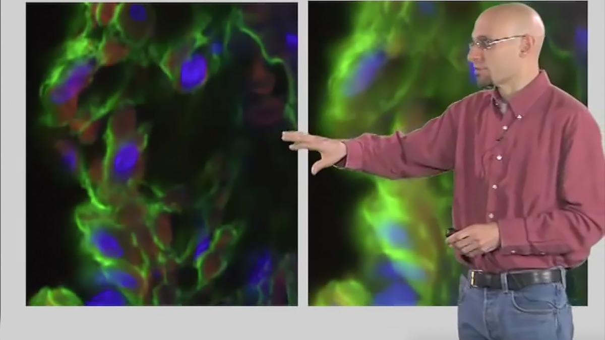
This talk introduces two-photon microscopy which uses intense pulsed lasers to image deep into biological samples. It can be used for imaging thick tissue specimens or even imaging inside of live animals.

Questions

  1. Explain why only red light is emitted through thin skin when a flashlight is shone on it.
  2. What is clearing and what is it used for?
  3. What are the advantages of using two infrared photons instead of one blue photon in two-photon microscopy to excite a green-light emitting molecule?
  4. Why does two-photon microscopy not require a pinhole (select all that apply)?
    1. Because the emitted light in two-photon microscopy does not scatter
    2. Because there is no out-focus light in two-photon microscopy
    3. Because two-photon microscopy gives rise to localized excitation
    4. None of the above
  5. What would be the result of using a pinhole in two-photon microscope?
    1. The detected image would include less background signal
    2. The detected image would be dimmer
    3. None of the above
  6. Do you agree or disagree with this statement? “In two-photon microscopy, one unique laser setting can be used to simultaneously excite both the Alexa Fluor 488 (green) and Alexa Fluor 568 (orange) dyes.” Explain why you agree or disagree.

Answers

View Answers
  1. Because biological materials absorb light in most wavelengths, except in the red color. Therefore, more red light is let through the skin than the other wavelengths
  2. Clearing removes scattering from biological samples. The solution uses vary and include improving “deep” imaging in thick fixed samples. The clearing solutions are either organic solvents, detergents, or urea.
  3. Using two photons instead of one will increase penetration depth; Because biological molecules do not usually absorb light in the red spectrum, using red photons to excite fluorescent markers can decrease the image background; In addition, two red photons used simultaneously can excite a fluorescent marker that is usually excited in the blue wavelength (in one-photon microscopy). This increases the range of fluorescent markers that can be used in thick sections; Last, because two-photon microscopy involves localized excitation, it does not involve pinholes.
  4. B and C
    1. Incorrect: The emitted light in two-photon microscopy does scatter, but it does not matter. Because only one point emits light (the focus of the light beam), the total emission is measured.
    2. Correct: Because there is no out-focus light in two-photon microscopy, the pinhole is not required in two-photon microscopy.
    3. Correct: Two-photon microscopy gives rise to localized excitation, which means that only the focus point receives sufficient light (from two photons) to be excited and emit light. Therefore, the excitation photons cannot produce sufficient light to excite markers, which means that there is no background light to filter through a pinhole.
  5. B
    1. Incorrect: The detected image would include less background signal. There is no background signal in two-photon microscopy
    2. Correct: The detected image would be dimmer. Because the total emitted light is measured in two-photon microscopy, and because the pinhole would filter out a significant portion of that emitted light, light would be lost if the pinhole were to be kept on.)
  6. Agree. Because two-photon microscopy uses two simultaneous photons of shorter wavelengths to excite dyes, the excitation spectrum of different dyes may overlap.

Speaker Bio

Kurt Thorn

Kurt Thorn

Kurt Thorn is an Assistant Professor of Biochemistry and Biophysics at UCSF and Director of the Nikon Imaging Center – a facility that provides cutting edge light microscopy equipment to UCSF researchers. Kurt can be followed on his blog at http://nic.ucsf.edu/blog/. Continue Reading

Playlist: Microscopy Series

  • Microscopy: Optical Sectioning and Confocal Microscopy (Kurt Thorn)
    Optical Sectioning and Confocal Microscopy
  • Light Sheet Sectioning (Ernst Stelzer)
    Light Sheet Sectioning
  • Hari Shroff
    Dual-View Inverted Selective Plane Illumination (diSPIM)
  • Total Internal Reflection Fluorescence (TIRF) Microscopy (Daniel Axelrod)
    Total Internal Reflection Fluorescence (TIRF) Microscopy

Reader Interactions

Comments

  1. D says

    May 15, 2020 at 11:39 pm

    Hi Kurt,

    Thank you for the awesome video which comprehensively explains the principles behind the two-photon microscopy imaging technique!

    Is there a download link for the slides used in this video?

    Thanks,
    D

    Reply
  2. Michal Jirak says

    December 21, 2020 at 2:25 am

    Great explanation, thank you.

    Reply

Leave a Reply Cancel reply

Your email address will not be published. Required fields are marked *

Primary Sidebar

Sign up for the Science Communication Lab education newsletter

  • Sign up Newsletter
  • This field is for validation purposes and should be left unchanged.

Privacy Policy

Help us keep bringing the world’s best biology to you!

Footer

Funders

NSF
NIGMS
Lasker
Rita Allen

Start Here

  • Talks for Everyone
  • Talks for Students
  • Talks for Research
  • Talks for Educators

Explore

  • Explore
  • All Playlists
  • All Speakers
  • All Talks

Talks By Topic

  • Biochemistry
  • Bioengineering
  • Biophysics
  • Cell Biology
  • Development and Stem Cells
  • Ecology
  • Genetics and Gene Regulation
  • Human Disease
  • Immunology
  • Microbiology
  • Neuroscience
  • Plant Biology
  • Techniques

Talks by Series

  • Bench to Bedside
  • Famous Discoveries
  • Great Questions
  • Share Your Research Series

Career

  • Professional Development
  • Career Exploration
  • NRMN Resources
  • Biomedical Workforce

Courses

  • Microscopy Series
  • Short Microscopy Series
  • Open edX Courses
  • Cell Biology Flipped Course
  • Engineering Life Flipped Course
  • Evolution Flipped Course

Educator

  • Educator Registration
  • Educator Resources
  • Log In

About Us

  • About Us
  • iBiology Team
  • Wonder Collaborative
  • Contact Us
  • Mission
  • Privacy Policy
  • SCL Financial Conflict of Interest Policy

This material is based upon work supported by the National Science Foundation and the National Institute of General Medical Sciences under Grant No. 2122350 and 1 R25 GM139147. Any opinion, finding, conclusion, or recommendation expressed in these videos are solely those of the speakers and do not necessarily represent the views of the Science Communication Lab/iBiology, the National Science Foundation, the National Institutes of Health, or other Science Communication Lab funders.

© 2023 - 2006 iBiology · All content under CC BY-NC-ND 3.0 license · Privacy Policy · Terms of Use · Usage Policy
 

Power by iBiology