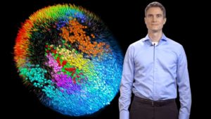
To better understand how an entire embryo develops from a single cell, Dr. Philipp Keller and colleagues developed a technique to image and quantitatively reconstruct mouse embryogenesis from gastrulation through early organogenesis at the single-cell level. Keller’s lab developed an adaptive light-sheet microscope to follow the mouse embryo for 48 hours while it’s developing its germ layers (mesoderm, endoderm, and ectoderm), early tissues, and organs. By combining long-term high-resolution imaging, computational, and statistical analyses, they generated a dynamic fate map of the embryo. These open-access resources aid in the understanding of the dynamic cell behaviors that allow for proper growth and development of the embryo.
View the full talk with additional resources on our website
Single-Cell Imaging: Imaging and Reconstructing Mouse Development at the Single-Cell Level
Dr. Philipp Keller describes the adaptive light-sheet microscope that his lab developed to image and quantitatively reconstruct mouse embryogenesis from gastrulation through early organogenesis at the single-cell level. (Talk recorded in December 2018)
Audience:
- Student
- Researcher
- Educators of H. School / Intro Undergrad
- Educators of Adv. Undergrad / Grad
Duration: 00:40:16
Speaker Bio
Philipp Keller