• Skip to primary navigation
  • Skip to main content
  • Skip to primary sidebar
  • Skip to footer

iBiology

Bringing the World's Best Biology to You

  • Start Here
    • Explore iBiology
    • All Playlists
    • All Speakers
    • All Talks
    • What’s new at iBiology?
  • Research Talks
    • Talks by Topic
      • Biochemistry
      • Bioengineering
      • Biophysics
      • Cell Biology
      • Development and Stem Cells
      • Ecology
      • Evolution
      • Genetics and Gene Regulation
      • Human Disease
      • Immunology
      • Microbiology
      • Neuroscience
      • Plant Biology
      • Techniques
      • Archive
    • Talks by Series
      • Bench to Bedside
      • Famous Discoveries
      • Great Unanswered Questions
      • Microscopy Series
      • Young Scientist Series
  • Stories
    • Background to Breakthrough
    • Interviews and Profiles
    • Science and Society
  • Courses
  • Careers
    • Professional Development Talks
    • Professional Development Courses
    • Career Exploration
    • NRMN Resources
    • Biomedical Workforce
  • Educator
  • About
    • Mission
    • iBiology Team
    • Board of Directors
    • Make a Donation
    • iBiology Funders and Partnerships
    • Contact Us
Home » Research Talks » Neuroscience

Visualizing Activity in the Brain

  • Duration: 10:39
  • Downloads
    • Hi-Res
    • Low-Res
  • Transcript

00:00:04.10 Hi. My name
00:00:08.11 is Adam Cohen. I am a professor
00:00:09.23 in the Departments of Chemistry
00:00:11.08 and Physics at Harvard.
00:00:12.13 Today I am going to talk
00:00:13.19 to you about
00:00:14.15 a huge unsolved problem
00:00:16.20 of how to visualize electrical activity
00:00:18.19 in the brain.
00:00:20.07 Now take a minute
00:00:21.17 and put your hand
00:00:22.03 on your head
00:00:23.04 and think about what you are touching.
00:00:24.05 Within a centimeter of your fingers
00:00:26.27 there is a piece of the most mysterious
00:00:29.03 and poorly understood matter
00:00:30.16 in the universe.
00:00:31.20 Here is a picture of your brain.
00:00:34.04 Now if we zoom in
00:00:35.12 by a factor of about 300
00:00:37.07 we see that this brain
00:00:39.08 is made up of neurons.
00:00:40.28 There is about a hundred billion of them,
00:00:43.05 and they are connected
00:00:44.08 to each other each by
00:00:45.18 about ten to the fourth,
00:00:46.17 10,000 connections.
00:00:48.07 If we zoom in again,
00:00:49.27 now by a factor of 10,000,
00:00:51.27 we see that each one
00:00:53.17 of these neurons is encased
00:00:54.27 in a lipid bilayer,
00:00:56.05 a little layer of grease,
00:00:57.23 which separates the inside of the cell
00:00:59.25 from the outside of the cell.
00:01:01.05 This bilayer is an electrical insulator
00:01:03.22 while the cytoplasm
00:01:04.19 and the extracellular medium
00:01:06.00 are conductors.
00:01:07.06 So you can think of it
00:01:07.27 a little bit like
00:01:09.08 a parallel plate capacitor.
00:01:11.13 Now there are many proteins
00:01:13.23 that sit in the membrane
00:01:15.00 and these proteins
00:01:16.12 can pass ions
00:01:18.13 across the membrane.
00:01:19.14 There are charge pumps,
00:01:20.17 which can generate a voltage,
00:01:22.02 and there are ion channels,
00:01:23.10 which can relieve this voltage.
00:01:24.15 These channels act
00:01:25.29 a little bit like resistors
00:01:26.25 except that they are non linear, so if
00:01:29.09 there is a change in the voltage,
00:01:30.15 they can have big changes
00:01:31.11 in the conductance
00:01:32.05 which can lead to these
00:01:33.15 electrical spikes
00:01:34.13 in the membrane voltage,
00:01:35.10 which we call
00:01:36.04 action potentials.
00:01:36.28 Every action potential
00:01:38.23 is about a tenth of a volt high
00:01:40.12 and lasts about
00:01:41.05 one one-thousandth of a second.
00:01:42.17 These are the currency
00:01:43.29 of the nervous system.
00:01:45.15 So every thought
00:01:46.17 and hope and dream
00:01:48.03 and feeling that we have
00:01:49.07 is encoded in these patterns
00:01:51.17 of electrical spikes
00:01:52.14 traveling through the
00:01:53.22 hundred billion neurons
00:01:54.17 in our brain.
00:01:55.03 That is all there is.
00:01:56.22 And so we'd really like
00:01:57.24 to see these action potentials
00:01:59.20 so that we can understand
00:02:01.10 what each neuron is doing
00:02:03.00 and also to look at
00:02:03.29 the global patterns of activity
00:02:06.00 and the interrelations
00:02:06.23 of the signals between
00:02:07.19 neurons to understand
00:02:08.28 how information
00:02:09.24 is being processed
00:02:10.21 within the nervous system.
00:02:12.04 So, basically this is a
00:02:14.25 voltage measurement problem.
00:02:15.23 Now traditionally this is measured,
00:02:18.19 as you might
00:02:18.29 measure the voltage
00:02:19.29 in any electrical circuit.
00:02:21.00 Electrophysiologists take a glass pipette
00:02:24.21 with a little electrode in it
00:02:26.19 and jam the pipette
00:02:27.13 into a cell and then there is
00:02:28.27 another electrode
00:02:29.18 out in the solution
00:02:31.11 and by hooking these up
00:02:31.24 to an amplifier
00:02:32.13 they can measure
00:02:33.11 the voltage difference.
00:02:34.09 And this works nicely,
00:02:35.07 but it only measures
00:02:36.17 one cell at a time.
00:02:38.20 It's extremely slow
00:02:40.03 and laborious to get these
00:02:41.12 electrodes into the cell,
00:02:42.23 and it doesn't give
00:02:44.06 you the global view of
00:02:45.24 what is going on in
00:02:46.19 a complex neural circuit.
00:02:48.18 Here is an artist's conception
00:02:50.24 of what we would really
00:02:51.26 like to do.
00:02:52.08 We'd like to just make these
00:02:53.18 neurons light up when they fire.
00:02:55.23 The problem is
00:02:56.26 that you can't see
00:02:57.25 the voltage in a neuron
00:02:59.15 anymore than you can see
00:03:00.28 the voltage in a telephone cord
00:03:02.11 when somebody is having
00:03:03.17 a telephone conversation.
00:03:04.17 We need some
00:03:05.22 contrast agent to convert
00:03:07.24 membrane voltage into light.
00:03:09.24 Now physicists and
00:03:12.06 chemists and biologists
00:03:13.18 have been working on trying
00:03:14.13 to invent contrast agent
00:03:16.09 since the late 1960s.
00:03:17.13 And this activity has really taken off
00:03:20.08 in the last few years.
00:03:21.27 Here are just a few
00:03:22.16 of the things
00:03:23.05 that people are working
00:03:23.28 on today. These are all from
00:03:25.16 within the last year or so.
00:03:26.15 People are trying to develop
00:03:28.23 voltage sensitive dyes.
00:03:30.02 So these are organic
00:03:31.07 small molecules which will
00:03:32.24 intercalate into the membrane,
00:03:34.28 and in some of these dyes,
00:03:36.10 illumination with light
00:03:38.03 generates an electronic excited state.
00:03:40.17 The charge distribution
00:03:42.03 in the excited state
00:03:43.10 is different than the
00:03:44.18 charge distribution in the ground state,
00:03:45.20 and so when there is a voltage
00:03:47.04 across the membrane
00:03:47.25 that voltage perturbs
00:03:49.26 the relative energy
00:03:51.05 of these two states
00:03:51.29 which can alter
00:03:52.25 the fluorescence properties
00:03:53.24 of the dye.
00:03:54.27 And so, this provides
00:03:55.22 a means to read out
00:03:56.18 membrane voltage with
00:03:57.16 fluorescence.
00:03:58.12 Other people are trying
00:03:59.29 to put semiconductor
00:04:01.00 nanocrystals
00:04:02.12 in lipid membranes.
00:04:03.13 These nanocrystals have
00:04:05.03 quantum-mechanical,
00:04:06.08 delocalized electronic states.
00:04:08.04 Membrane voltage can again
00:04:09.14 shift the relative energies
00:04:10.23 of these states
00:04:12.00 and adjust again the
00:04:13.06 fluorescent properties
00:04:13.29 of these crystals.
00:04:15.02 Other people are working
00:04:16.21 with objects called
00:04:18.00 nitrogen vacancies in diamonds.
00:04:19.24 These are tiny quantum
00:04:21.20 mechanical defects
00:04:22.22 in diamonds
00:04:23.23 and people think that if you
00:04:25.06 get a small enough
00:04:26.03 diamond nanocrystal
00:04:27.24 and you stick it into a membrane
00:04:29.22 the fluorescent properties
00:04:30.28 of these nitrogen vacancies
00:04:32.07 will again be sensitive
00:04:33.11 to membrane voltage.
00:04:34.05 Now I want to emphasize
00:04:36.00 these are all interesting
00:04:37.26 prospective technologies,
00:04:39.02 but none of these
00:04:40.02 have solved the problem
00:04:41.02 that I posed at the beginning yet.
00:04:42.19 In all of these technologies
00:04:45.08 the sensor is sitting
00:04:47.04 in the membrane.
00:04:47.23 And this is because
00:04:48.26 to measure a voltage difference,
00:04:49.26 you need to be connected
00:04:50.26 both to the inside of the cell
00:04:52.10 and the outside of the cell.
00:04:53.09 Other people, including myself,
00:04:56.05 are working on voltage indicators
00:04:59.09 based on fluorescent
00:05:00.18 transmembrane proteins.
00:05:02.07 The idea is to have a protein
00:05:04.05 which sits in a membrane
00:05:05.13 and to somehow
00:05:06.04 make its fluorescence
00:05:07.07 sensitive to the membrane voltage.
00:05:08.22 I'll tell you just a
00:05:09.16 little bit about one
00:05:10.14 indicator which we have been
00:05:11.17 developing in my lab.
00:05:12.17 The story starts here.
00:05:15.09 This is a picture of the Dead Sea.
00:05:17.11 There is a micro-organism that lives
00:05:19.19 in the water here that produces
00:05:21.21 a transmembrane protein
00:05:22.28 called archaerhodopsin-3.
00:05:24.17 This protein absorbs sunlight
00:05:27.08 and uses that energy
00:05:29.05 to pump a proton, a charge,
00:05:30.17 from inside the cell
00:05:31.24 to outside the cell.
00:05:32.29 This generates a membrane voltage,
00:05:35.16 which this creature uses
00:05:36.02 to power its metabolism.
00:05:37.10 So a few years ago
00:05:39.24 I said to myself
00:05:40.19 I wonder if we
00:05:41.13 can run this thing
00:05:42.10 in reverse.
00:05:43.07 Can we use a change
00:05:44.26 in membrane voltage
00:05:46.02 to induce a conformational
00:05:47.22 change in this protein
00:05:48.22 which will lead to a fluorescence
00:05:51.00 signal that we can see?
00:05:52.02 Just like this. Right.
00:05:54.15 And you see this works
00:05:55.20 in Powerpoint,
00:05:56.06 so that is very encouraging.
00:05:56.24 After several years
00:05:58.28 we engineered
00:05:59.11 a protein which
00:06:00.00 had these attributes.
00:06:00.21 And here is a picture
00:06:03.18 of a human tumor cell
00:06:05.16 where we have introduced
00:06:06.15 a gene for this protein
00:06:08.14 and we are using a little patch pipette,
00:06:11.09 which you can't see,
00:06:11.25 to induce steps of voltage
00:06:13.21 between plus and minus
00:06:14.14 a tenth of a volt,
00:06:15.19 and we are looking at the fluorescence
00:06:16.19 of the protein.
00:06:18.17 So here you can see
00:06:20.15 that as the voltage goes up,
00:06:21.23 the protein gets bright,
00:06:22.21 and as the voltage goes down
00:06:23.29 the protein gets dim.
00:06:25.06 So this is a
00:06:26.05 genetically encoded fluorescent
00:06:27.28 reporter of membrane voltage,
00:06:29.05 and it turns out
00:06:30.03 that it can be quite fast
00:06:30.28 and sensitive
00:06:31.21 but also very dim,
00:06:32.24 which makes it hard to measure.
00:06:34.17 We have now developed technology
00:06:35.27 to take these proteins
00:06:37.08 and to express them
00:06:38.27 in neurons in a Petri dish.
00:06:40.17 So the neurons become fluorescent
00:06:42.04 and their fluorescence
00:06:43.07 becomes sensitive
00:06:44.05 to the voltage
00:06:44.27 in the membrane.
00:06:45.05 We are also expressing
00:06:46.21 another protein in these cells
00:06:49.17 which renders them
00:06:50.11 sensitive to blue light.
00:06:52.17 This is a light gated
00:06:53.24 ion channel
00:06:54.25 and targeting a
00:06:56.11 flash of blue light
00:06:57.07 to the cell body
00:06:57.22 will trigger the
00:06:58.25 neuron to fire.
00:06:59.24 We have developed
00:07:01.00 computational techniques
00:07:02.07 to infer the propagation
00:07:04.03 of these electrical impulses
00:07:05.25 at an extremely high time resolution.
00:07:08.01 Here I will show you a movie
00:07:09.09 at a one hundred thousand
00:07:10.15 frames per second
00:07:11.14 of this optically induced
00:07:13.01 action potential
00:07:14.02 propagating through a neuron.
00:07:15.10 And you can really see
00:07:18.17 how these signals
00:07:19.15 propagate through a cell now,
00:07:21.08 but this is a neuron in culture.
00:07:24.03 We have taken a rat brain
00:07:24.20 and we've mushed it up
00:07:25.19 and we spread out
00:07:26.14 the neurons in a dish.
00:07:27.21 We'd really like to do this
00:07:29.11 in an intact brain,
00:07:31.00 but neither we
00:07:31.21 nor anybody else on the planet
00:07:33.00 knows how to do that right now.
00:07:34.22 There are several challenges to this,
00:07:36.21 and I'll tell you about them.
00:07:37.25 So one of the
00:07:41.27 problems we face
00:07:42.26 is that the brain
00:07:44.07 is not transparent.
00:07:45.21 Here is a picture
00:07:46.20 of a mouse brain.
00:07:48.23 And you can see
00:07:50.02 that it scatters light
00:07:51.21 very strongly.
00:07:52.02 It's about the size of my pinky
00:07:54.15 but light scatters
00:07:54.28 over a distance of
00:07:55.23 less than a tenth of a millimeter.
00:07:57.07 And so it is an
00:07:58.28 unsolved challenge how to
00:08:00.06 visualize what is going on
00:08:01.25 inside of this chunk of tissue.
00:08:03.24 We need to come up with new kinds
00:08:05.01 of optical or electrical tricks.
00:08:07.14 Another challenge
00:08:09.00 is that in every
00:08:10.07 mouse brain there is about
00:08:11.17 75 million neurons.
00:08:13.02 To record from these
00:08:14.20 at a thousand frames per second,
00:08:16.03 which you need to detect
00:08:17.16 action potentials
00:08:18.06 would lead to a
00:08:19.05 data rate of about
00:08:20.25 75 gigabytes a second.
00:08:22.15 We don't know how
00:08:24.08 to store this data, let alone
00:08:25.23 what to do with it
00:08:26.21 if we did have it.
00:08:28.02 So this brings me to
00:08:29.21 another unsolved challenge,
00:08:31.00 which is the interpretation problem.
00:08:33.23 So even if we had the best
00:08:36.02 technology
00:08:36.15 imaginable for visualizing
00:08:38.11 electrical activity in the brain,
00:08:39.26 how do we
00:08:40.24 interpret it?
00:08:42.00 We'd like to relate
00:08:43.08 the electrical activity
00:08:45.02 to some statement about
00:08:46.27 the underlying biology
00:08:48.05 of the cells.
00:08:48.20 What are the ions channels
00:08:49.24 that are active?
00:08:50.19 What's their state
00:08:51.07 of regulation?
00:08:52.05 What's their distribution?
00:08:53.09 What's the metabolic
00:08:54.17 state of the cell?
00:08:55.26 What's happening
00:08:56.13 to its energy supply?
00:08:57.20 We all experience that
00:08:58.24 as we go through our
00:09:00.11 day to night cycle
00:09:01.24 the function of our neurons
00:09:02.16 changes tremendously,
00:09:03.28 whether we are asleep or awake,
00:09:05.08 tired or not tired,
00:09:06.06 hungry or not hungry.
00:09:07.15 And those changes
00:09:08.12 are not mediated
00:09:09.18 by structural changes
00:09:10.11 in the brain but
00:09:11.13 by changes in presumably
00:09:13.06 the metabolic state
00:09:14.13 of our neurons.
00:09:15.13 And then finally we'd like
00:09:17.09 to infer from patterns of activity
00:09:20.02 how the neurons are connected
00:09:21.21 to each other.
00:09:22.12 To figure out who is calling
00:09:23.07 the shots and
00:09:24.07 who is controlling who
00:09:25.02 and how they
00:09:25.26 integrate and process information.
00:09:28.03 To illustrate this,
00:09:28.28 here is a
00:09:29.23 movie of some neurons.
00:09:31.27 This was a movie
00:09:32.15 taken in my lab.
00:09:33.04 These are neurons in a dish,
00:09:34.14 not in an intact brain
00:09:35.28 where we will look at
00:09:37.00 their firing patterns.
00:09:38.00 This is a movie at
00:09:39.11 about five hundred
00:09:39.26 frames per second.
00:09:40.15 The neurons have been
00:09:41.08 artificially colored
00:09:42.06 just so you can tell them apart.
00:09:43.28 And we can zoom in
00:09:45.28 on anyone of these cells.
00:09:47.23 and look at its firing pattern,
00:09:49.01 and we really don't
00:09:50.10 know how to interpret
00:09:51.22 these firing patterns
00:09:52.20 in terms of the ion channels,
00:09:54.14 the energetics,
00:09:55.12 and the connectivity of these cells.
00:09:57.23 So there is tremendous scope
00:10:00.18 over the coming years
00:10:01.15 to combine the efforts
00:10:03.06 of physicists, chemists, and biologists
00:10:04.29 to develop technology
00:10:07.10 which will let us visualize
00:10:08.27 the activity
00:10:09.27 in this little chunk of matter
00:10:12.02 that is right between
00:10:12.22 all of our ears.
00:10:14.14 Thanks.
00:10:15.03

This Talk
Speaker: Adam Cohen
Audience:
  • Educators of Adv. Undergrad / Grad
  • Researcher
  • Educators
Recorded: July 2014
More Talks in Great Unanswered Questions
  • Julie Theriot
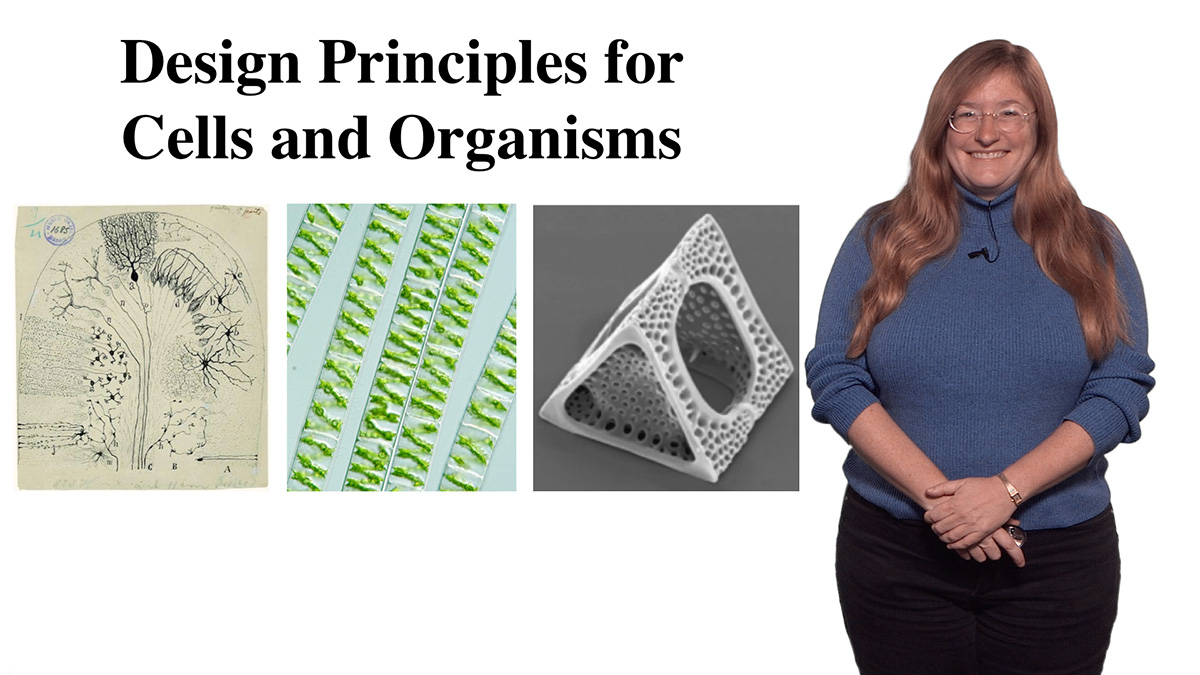
    Cellular Structures: Discovering Design Principles for Cells and Organisms
  • Jennifer Lippincott-Schwartz
    How Do Lipids and Cholesterol Regulate Trafficking?
  • Michael Rosen
    Mechanisms of Cell Organization on Micron Length Scales
All Talks in Great Unanswered Questions
Share

Talk Overview

The pattern of electrical signals propagated through neuronal networks determines brain function. Adam Cohen examines the possibility of visualizing the brain function inside an intact brain using fluorescent transmembrane proteins that are sensitive to voltage. Cohen discusses the barriers to this approach, something he predicts scientists from many disciplines will eventually overcome.

Speaker Bio

Adam Cohen

Adam Cohen

Adam Cohen is Professor in the Departments of Chemistry and Physics at Harvard University and Investigator of the Howard Hughes Medical Institute. He develops biological tools and analytical approaches to investigate the behaviors of molecules and cells in vitro and in vivo. His lab merges protein engineering, optics, and physics, among other disciplines, on a… Continue Reading
More Talks in Neuroscience

  • Ricardo Dolmetsch
    Neurodevelopmental disease: Drug Discovery in Neuroscience
  • Roger Hanlon
    Rapid Adaptive Camouflage and Signaling in Cephalopods
  • Torsten Wiesel
    Exploring the Visual Brain
  • How Flies Fly: Lift: Michael Dickinson
    How Flies Fly

Reader Interactions

Leave a Reply Cancel reply

Your email address will not be published. Required fields are marked *

Primary Sidebar

Like our Website?

You'll love our newsletter...

  • Sign up for:
    • Exclusive iBiology content
    • 10 Must See Biology Videos
    • Talks by 20+ Nobel Winners
    • Our young Scientist Survival Toolkit
    • Talks by trailblazing Women in Biology
    • Updates on exciting iBiology projects in development
    Tyler Allen
  • This field is for validation purposes and should be left unchanged.

Privacy Policy

Help us keep bringing the world’s best biology to you!

Footer

Funders

NSF Logo
Lasker Logo
NIGMS Logo

Partners

Start Here

  • Talks for Everyone
  • Talks for Students
  • Talks for Research
  • Talks for Educators

Explore

  • Explore
  • All Playlists
  • All Speakers
  • All Talks

Talks By Topic

  • Biochemistry
  • Bioengineering
  • Biophysics
  • Cell Biology
  • Development and Stem Cells
  • Ecology
  • Genetics and Gene Regulation
  • Human Disease
  • Immunology
  • Microbiology
  • Neuroscience
  • Plant Biology
  • Techniques

Talks by Series

  • Bench to Bedside
  • Famous Discoveries
  • Great Questions
  • Share Your Research Series

Career

  • Professional Development
  • Career Exploration
  • NRMN Resources
  • Biomedical Workforce

Courses

  • Microscopy Series
  • Short Microscopy Series
  • Open edX Courses
  • Cell Biology Flipped Course
  • Engineering Life Flipped Course
  • Evolution Flipped Course

Educator

  • Educator Registration
  • Educator Resources
  • Log In

About Us

  • About Us
  • iBiology Team
  • Wonder Collaborative
  • Contact Us
  • Make a Donation
  • Mission
  • Privacy Policy

This material is based upon work supported by the National Science Foundation and the National Institute of General Medical Sciences under Grant No. MCB-1052331. Any opinion, finding, conclusion, or recommendation expressed in these videos are solely those of the speaker and do not necessarily represent the views of iBiology, the National Science Foundation, the National Institutes of Health, or other iBiology funders.

© 2021 - 2006 iBiology · All content under CC BY-NC-ND 3.0 license · Privacy Policy · Terms of Use · Usage Policy
 

Power by iBiology