• Skip to primary navigation
  • Skip to main content
  • Skip to primary sidebar
  • Skip to footer

iBiology

Bringing the World's Best Biology to You

  • Start Here
    • Explore iBiology
    • All Playlists
    • All Speakers
    • All Talks
    • What’s new at iBiology?
  • Research Talks
    • Talks by Topic
      • Biochemistry
      • Bioengineering
      • Biophysics
      • Cell Biology
      • Development and Stem Cells
      • Ecology
      • Evolution
      • Genetics and Gene Regulation
      • Human Disease
      • Immunology
      • Microbiology
      • Neuroscience
      • Plant Biology
      • Techniques
      • Archive
    • Talks by Series
      • Bench to Bedside
      • Famous Discoveries
      • Great Unanswered Questions
      • Microscopy Series
      • Share Your Research Series
  • Stories
    • Background to Breakthrough
    • Interviews and Profiles
    • Science and Society
  • Courses
  • Careers
    • Professional Development Talks
    • Professional Development Courses
    • Career Exploration
    • NRMN Resources
    • Biomedical Workforce
  • Educators
  • About
    • Mission
    • Commitment to Inclusion
    • iBiology Team
    • Board of Directors
    • iBiology Funders and Partnerships
    • Contact Us
  • Donate
Home » Research Talks » Development and Stem Cells

Molecular Mechanisms that Drive Regeneration

  • Duration: 27:18
  • Downloads
    • Hi-Res
    • Low-Res
  • Subtitles
    • English
  • Transcript

00:00:07.06 My name is Peter Reddien, and I'm an Associated Professor of Biology at MIT,
00:00:12.01 a member of the Whitehead Institute,
00:00:13.27 and an Early Career Scientist of the Howard Hughes Medical Institute.
00:00:16.26 Today, I'm going to be talking to you about a manuscript published in Science magazine
00:00:22.05 entitled Polarized notum Activation at Wounds Inhibits Wnt function to Promote
00:00:27.18 Planarian Head Regeneration.
00:00:28.26 So, this was published by Christian Petersen and myself, and it has been focused on
00:00:34.06 in an educational website at Science called Science in the Classroom.org.
00:00:40.13 The focus of this manuscript is on the question, how do regenerative animals determine
00:00:45.26 what part of their body is missing, such that they initiate the correct regeneration programs
00:00:50.11 to replace those missing body parts?
00:00:53.17 The ability of animals to regenerate is widespread in the animal kingdom.
00:00:58.00 Shown here are many familiar examples, such as earthworms that can regenerate following
00:01:01.18 being pulled in half, starfish, salamanders, cephalopods, planarians,
00:01:07.21 and there are many other examples of animals that can replace missing body parts through regeneration.
00:01:13.08 In fact, regeneration is one of the great mysteries of biology.
00:01:17.03 Biologists have long been captivated with this phenomenon of regeneration.
00:01:20.08 But it has only been in recent years that we've begun to elucidate some of
00:01:24.17 the key molecular mechanisms of the process.
00:01:29.12 Observing these feats of regeneration in these animals elicits the question,
00:01:34.00 how does it work in these animals?
00:01:35.16 How are they so good at this process?
00:01:37.20 And why are we less adept at regenerating missing body parts?
00:01:41.11 To get insight into this question of how regeneration works, it is important to have model systems
00:01:48.08 that allow us to investigate the cellular and molecular mechanisms that underlie the process.
00:01:54.12 I became captivated with this problem when I was late in my graduate career.
00:01:59.17 I had experienced the power of simple organisms that could powerfully and readily be studied
00:02:06.07 in the laboratory for elucidating generalizable principles of biology.
00:02:11.13 And I was interested in studying regeneration in simple organisms that could be
00:02:16.15 readily studied in the laboratory.
00:02:18.10 A number of things are important for this.
00:02:21.20 One is the ability of the organism to regenerate robustly; the ability to regenerate rapidly
00:02:28.05 for performing a large number of experiments;
00:02:31.15 the ability to culture the animal in the laboratory in large numbers;
00:02:34.23 and ideally, mechanisms or methods could be devised for the study of cells
00:02:38.22 underlying regeneration and the genes controlling regeneration.
00:02:44.08 Planarians, in the past 10-15 years, have emerged as a new and powerful molecular system
00:02:50.02 for studies of regeneration.
00:02:52.06 This is a planarian.
00:02:53.27 Planarians are easily recognized by their sort of cartoon-like crossed eyes.
00:02:57.26 They have a pharynx in the middle of the animal.
00:03:00.04 And they are a classic model for studies of regeneration.
00:03:03.20 They are capable of regenerating entire new heads following amputation;
00:03:07.27 or entire new tails;
00:03:10.19 or an entire individual, a whole animal, from a small fragment of the body.
00:03:16.19 Regeneration is accomplished in part by the process of blastema formation.
00:03:21.21 Shown here is an animal in which its head has been amputated.
00:03:25.17 And this animal is regrowing a new head.
00:03:28.04 The process takes about a week.
00:03:30.14 The blastema is seen as an unpigmented outgrowth at the wound site.
00:03:35.03 It appears within 2-3 days.
00:03:37.06 And within this blastema, some of the missing tissues will differentiate such as,
00:03:41.10 in this case, eyes and brain.
00:03:45.17 Planarians have a complex internal anatomy, including eyes, brain, and other sensory systems.
00:03:50.17 They're surrounded by an epidermis.
00:03:52.24 They have a musculature for twisting and turning.
00:03:55.17 They have a highly branched intestinal system for delivering nutrients.
00:03:59.18 They have a pharynx in the middle of their body for feeding.
00:04:02.13 They have an excretory, kidney-like system.
00:04:04.21 And there are dozens of other cell types that have been recognized.
00:04:08.09 Because a tiny fragment of the body can regenerate an entire organism, there must exist mechanisms
00:04:13.27 in the adult for the production of all of these cell types, organs, and tissue patterns.
00:04:19.21 How that happens is the focus of the research in my laboratory.
00:04:23.25 How do we investigate this process?
00:04:27.06 Well, it is possible to study the location and timing of expression of any gene desired
00:04:33.28 in the process of regeneration.
00:04:36.11 Let me illustrate with a brief example how this works.
00:04:40.11 During the development of animals, many different cell types emerge
00:04:44.09 -- in this hypothetical example, cell types A and B --
00:04:48.04 and these cells will have the same genome present within them.
00:04:51.15 And they are made different from one another not by the presence of the genes in the genome
00:04:56.06 but by how those genes are used.
00:04:58.16 For example, some genes will be on in cell type A, like gene 1, here, and off in cell type B.
00:05:05.03 Other genes might be on, like gene 2 in cell type B but off in cell type A.
00:05:11.14 Other genes might be on in both cell types.
00:05:14.22 Now, these genes are going to be encoding mRNA that corresponds a nucleic acid sequence
00:05:22.03 to the sequence of that gene.
00:05:24.13 This mRNA then encodes a protein that carries out the function encoded by that gene.
00:05:31.07 It is possible to detect, experimentally, the mRNA for gene 1,
00:05:37.05 or the protein encoded by gene 1, within the planarian.
00:05:41.25 For example, we can label the mRNA for gene 1 or the protein encoded by gene 1 with
00:05:48.14 a fluorescent RNA probe for the mRNA or with the fluorescent antibody for the protein.
00:05:56.24 In this example, imagine we have an RNA probe for the mRNA for gene 1 that has been
00:06:02.26 chemically treated such that it will fluoresce red.
00:06:07.25 In this case, then, cell type A would turn red in the experiment.
00:06:11.11 Now imagine we add an RNA probe that will fluoresce green, and this RNA probe is specific
00:06:17.28 to the mRNA for gene 2.
00:06:19.26 Then, cell type B will turn green.
00:06:23.10 And in this way, we can visualize any cell desired in the intact planarian and observe
00:06:29.01 its regeneration.
00:06:30.22 For example, shown here is the planarian excretory system, its kidney-like protonephridia.
00:06:37.12 Here's the musculature of an entire animal, from head to tail.
00:06:42.10 These are the eyes of planarians.
00:06:44.10 They have two cell types: photosensitive neurons and shading pigment cells.
00:06:50.16 The nervous system, the intestine, and any other cell type.
00:06:55.00 Shown here is a particularly important cell type for regeneration.
00:06:59.04 In red are the cells called neoblasts.
00:07:02.13 And the neoblasts are the dividing cells of the adult planarian.
00:07:06.19 And they are the cells responsible for the production of all of the missing cell types
00:07:10.28 during regeneration.
00:07:13.15 Within the neoblast population are cells that are called pluripotent, and they are stem cells.
00:07:19.09 They are capable of dividing and making more neoblasts, and all of the cells of
00:07:23.26 the adult tissue of the planarian.
00:07:25.05 In this talk today, I'm focusing not on the neoblasts and how they work but the instructions.
00:07:32.12 What tells the neoblasts what to do following injury?
00:07:36.06 What are the instructions that specify what part of their body should be regenerated?
00:07:42.10 To investigate genes, we can not only look at where they are active but we can perturb
00:07:47.26 their function.
00:07:48.26 The goal is to take planarian genes and break them, and ask what happens to
00:07:54.11 the process of regeneration when we break a particular gene.
00:07:57.18 We aim to infer what that gene normally does by what step in regeneration goes awry
00:08:03.26 in the absence of function for that gene.
00:08:06.27 To do this, we need a parts list.
00:08:08.21 We need our hands on the complete set of genes in the planarian genome.
00:08:13.21 And this is now possible.
00:08:15.08 So, the planarian genome has been entirely sequenced, and I show, here,
00:08:18.28 a couple of key figures in this process.
00:08:20.23 So, I started my work on planarians in the early 2000s
00:08:26.05 as a postdoc in the lab of Alejandro Sanchez Alvarado, photographed here.
00:08:31.07 And together with Phil Newmark, we set out to attempt to sequence the planarian genome.
00:08:37.12 And this is a picture from Washington University at their genome sequencing center,
00:08:41.04 where this project was completed.
00:08:42.28 Now, Phil Newmark and Alejandro were working in the late '90s at the Carnegie Institute.
00:08:49.24 And a neighboring lab at this Institute was the lab of Andy Fire, who was involved in
00:08:54.11 the discovery of a mechanism called RNA interference, which can be used to break the function of genes.
00:09:01.04 And Alejandro and Phil found that this could work in planarians.
00:09:06.06 Here's the idea.
00:09:07.06 So, RNA interference, or RNAi, involves the use of double-stranded RNA that is...
00:09:14.22 corresponds in sequence specifically to a particular gene.
00:09:19.12 Through the mechanism of RNA interference, the double-stranded RNA will destroy the mRNA
00:09:24.16 encoded by a particular gene, thus blocking its ability to make protein and carry out
00:09:28.18 its function.
00:09:29.25 And in this way, we can choose any gene desired and inhibit it, and ask what happens to regeneration.
00:09:38.08 One way the double-stranded RNAs can be delivered for RNA interference is by using bacteria
00:09:44.13 as a factory to produce the double-stranded RNAs desired.
00:09:48.11 So, here's an example cartoon E coli bacterium, producing,
00:09:53.24 in this case, planarian double-stranded RNA sequence.
00:09:57.22 We then feed these bacteria to planaria.
00:10:00.06 Now, planaria don't like to eat bacteria, so we make them tasty to the animals by
00:10:05.03 mixing the bacteria with their normal laboratory food of blended liver.
00:10:09.12 Planarians like to eat this concoction.
00:10:11.10 The bacteria lyse in the intestine, releasing the double-stranded RNA cargo.
00:10:15.14 It spreads throughout the tissue of the animal.
00:10:18.04 And this breaks the function of the gene by inhibiting the mRNA for the gene in all of
00:10:22.24 the tissue types of the animal that we have looked at.
00:10:26.06 Now, early in my work on planarian regeneration,
00:10:30.05 while working together with Alejandro Sanchez Alvarado,
00:10:33.19 I set out to try to develop methods for performing what are called RNAi screens.
00:10:38.02 The idea is to take a large number of genes and inhibit them one at a time and systematically,
00:10:44.25 going through hundreds to over a thousand genes.
00:10:48.11 And this worked incredibly well.
00:10:49.28 We found a large number of defects in regeneration following inhibition of a number of different genes.
00:10:55.24 I show some examples here.
00:10:57.09 Here's an example where, following inhibition of a gene, regeneration failed.
00:11:01.15 Here's another example where regeneration occurred, but the blastema had
00:11:06.12 an abnormal shape, or morphology.
00:11:08.19 Here's an example of a growth coming out of the animal following inhibition of a particular gene.
00:11:14.04 An animal that would stretch.
00:11:15.15 Here's one that would bloat or blister.
00:11:17.28 And there are many other interest defects in the process of regeneration that can be studied.
00:11:22.17 Now, I'd like to point out that the majority of genes associated with these defects,
00:11:28.11 what are called phenotypes, are evolutionarily conserved,
00:11:32.07 meaning counterparts to these genes are found in animals throughout the animal kingdom, including in us.
00:11:38.08 So, we can ascribe functions for these genes by using planarians as a model system
00:11:44.13 to study this stem cell and regenerative biology that they so readily enable us to investigate in the lab.
00:11:53.16 Planarians have been investigated for well over a century.
00:11:57.02 And some of the first systematic surgical manipulations and observations of
00:12:01.15 planarian regeneration were performed by TH Morgan in the late 1800s and early 1900s.
00:12:08.13 And this TH Morgan is the founder of Drosophila genetics, a famous figure in the field of genetics.
00:12:14.20 He was captivated with the ability of planarians to regenerate their bodies from small fragments.
00:12:19.26 And he posed a problem about a century ago, which he referred to as regeneration polarity.
00:12:25.17 Which is a classic problem for thinking about tissue polarity and decision making
00:12:30.09 in regeneration at wounds.
00:12:32.17 You can think about the problem of regeneration polarity with the following thought experiment.
00:12:38.19 Imagine two hypothetical amputation planes: I and II.
00:12:42.25 Following amputation at plane I, you can produce an animal in which the cells circled in green
00:12:48.26 become involved in head regeneration.
00:12:51.13 Similarly, you can produce an animal following amputation at plane II,
00:12:55.25 where the same cells become involved in tail regeneration.
00:12:59.20 How did they know?
00:13:00.26 How did they figure out whether they were missing their head or their tail?
00:13:07.08 To investigate this process, I must introduce a key figure for this story.
00:13:12.19 Chris Petersen was the first postdoc to join my lab, in 2006 at MIT.
00:13:18.14 And we were interested in how planarians repattern their tissues during regeneration.
00:13:25.04 Chris was performing an RNAi screen in which he was systematically inhibiting genes
00:13:30.06 and looking at what happened in regeneration.
00:13:33.03 He was looking in particular at this middle piece.
00:13:36.24 This middle piece is regenerating a head at one wound site and a tail at the other wound site.
00:13:42.21 And the key result from the screen can be summarized with a single image.
00:13:49.05 When the gene beta-catenin was inhibited, animals regenerated, instead of a tail,
00:13:55.13 a second head, producing a two-headed animal, with both heads fighting to pull
00:13:59.16 the same body in opposite directions.
00:14:01.24 This was a spectacular phenotype for us.
00:14:04.13 We instantly knew this was a really critical clue to figuring out this process of
00:14:10.00 regeneration polarity, how these wounds know which way they're facing.
00:14:17.08 Now, I'll point out that we published this paper in 2008.
00:14:21.14 And I reference here two other manuscripts from the Sanchez Alvarado lab and the Salo lab,
00:14:25.07 which also provide nice analyses of this phenotype in planarians.
00:14:33.08 We found that this gene, beta-catenin, is working in regeneration to inhibit head regeneration
00:14:40.00 at wound sites.
00:14:41.21 And I think this concept is most dramatically illustrated with this experiment.
00:14:46.02 So, in this experiment, we inhibited beta-catenin with RNAi.
00:14:50.16 And then we removed the tail and made a series of incisions in the side of the body.
00:14:57.11 What happened following these incisions was the regeneration of this six-headed animal
00:15:02.19 you see here, which at the time, for me, was the most spectacular phenotype I had ever seen.
00:15:08.26 So, this six-headed animal illustrates this concept that beta-catenin is required at wounds
00:15:14.14 to prevent head regeneration.
00:15:18.25 How does it work?
00:15:21.19 Beta-catenin is a well characterized mediator of a process called Wnt signaling.
00:15:27.11 Beta-catenin exists in all animals, as does Wnt signaling, including in us.
00:15:32.09 And Wnt signaling is a means of cell-cell communication.
00:15:36.24 One cell will produce a secreted protein called Wnt.
00:15:41.02 Wnt can then interact with a second cell by binding to a different protein at the surface of that cell
00:15:48.10 called Frizzled.
00:15:49.17 Then, through a complex biochemical mechanism, Frizzled can lead to the activation of
00:15:55.20 the beta-catenin protein.
00:15:58.05 Active beta-catenin then will orchestrate gene expression in this recipient cell,
00:16:04.25 controlling which genes are on and off.
00:16:09.09 To investigate whether Wnt signaling is a key process for controlling regeneration polarity,
00:16:15.28 we looked at planarian Wnt genes.
00:16:18.10 And we found that a number of them are expressed in a regional way, constitutively,
00:16:23.25 in the adult body.
00:16:25.17 These patterns were quite striking to us, as we knew that beta-catenin was
00:16:30.28 required for tail regeneration, and when we look at the Wnt genes, we see that they're expressed
00:16:35.10 in the tail.
00:16:37.02 This Wnt gene, wnt1, is expressed in a handful of cells, just at the tip of the tail.
00:16:42.00 And here's two other Wnt genes that are expressed in a posterior-to-anterior gradient, transcriptionally,
00:16:48.04 at the mRNA level, extending from the tip of the tail up towards the head.
00:16:53.08 Now, from systematic investigation of these genes, we found that one of these Wnt genes
00:17:00.01 is a key gene for the process of regeneration polarity.
00:17:03.21 And that is the gene wnt1. wnt1 is the Wnt gene expressed just in a few cells
00:17:12.09 at the tip of the tail.
00:17:13.27 We looked at what happens to wnt1 expression during regeneration.
00:17:20.16 This is a wound facing the old tail, or posterior-facing.
00:17:26.08 And what is seen is that wnt1 expression, in purple, ramps up
00:17:30.20 at these posterior-facing wound sites within hours -- shown here is 12 hours -- of injury.
00:17:35.21 Each one of these purple dots is a single cell.
00:17:39.26 This makes sense.
00:17:40.27 wnt1 we found to be required for tail regeneration instead of head regeneration.
00:17:45.27 And we see it coming on during tail regeneration at wound sites.
00:17:49.23 To our surprise, we found that wnt1 was also expressed at head-facing wounds,
00:17:57.13 or what are called anterior-facing wounds.
00:17:59.28 And that's seen in this fragment here.
00:18:02.25 In fact, because we saw it was at both of these types of wounds,
00:18:06.23 we wondered if it would be activated at all types of wounds, and in fact found that to be the case.
00:18:12.28 Any type of injury we have inflicted upon these animals that incises the epidermis
00:18:18.15 activates the expression, at that wound site, of wnt1,
00:18:22.06 suggesting wnt1 is generically activated by wounding.
00:18:27.25 This raised a puzzle to us.
00:18:30.14 If wnt1 is active at posterior-facing wounds, tail-facing wounds, to promote a tail to regenerate,
00:18:38.25 what happens at the head-facing wounds, or anterior-facing wounds?
00:18:42.23 The suggestion would be that wnt1 is not active there for promoting a tail.
00:18:47.12 How could that be accomplished?
00:18:51.11 We found a key gene for this process.
00:18:54.00 And that gene is called notum.
00:18:56.26 notum has a very striking expression pattern.
00:19:01.12 Opposite to wnt1, notum is expressed at the tip of the head of planarians.
00:19:08.00 notum is a protein that is broadly found in the animal kingdom
00:19:12.00 but has not been extensively studied.
00:19:14.15 It is found... and was originally found and described for its roles in fruit flies, Drosophila.
00:19:22.12 When we looked at notum, we saw this striking expression pattern at the tip of the head.
00:19:26.16 And given its known biochemical function, we thought this was going to be a good suspect
00:19:31.04 for being involved in regeneration polarity.
00:19:34.08 Biochemically, what notum does is act outside of cells.
00:19:39.08 It's a secreted protein.
00:19:40.23 And it codes an enzyme called a hydrolase.
00:19:43.15 And it can act to cleave certain cell surface proteins off the surface.
00:19:48.14 This is an example.
00:19:49.14 So, a glypican is a cell surface protein that can help some signaling proteins, like Wnts,
00:19:56.20 bind to the cell surface and interact with their receptor.
00:20:00.24 notum could cleave the glypican off the surface of the cell.
00:20:04.20 This cleaved glypican could still interact with some signaling ligands, in principle,
00:20:10.10 and then might inhibit their ability to interact with their receptor.
00:20:15.07 Thus, notum is a candidate inhibitor of Wnt signaling.
00:20:21.11 When we looked at notum expression during regeneration, what we saw is that notum
00:20:26.00 is induced to be expressed at wounds, and rapidly following wounding.
00:20:30.27 But interestingly, its expression was preferential for head-facing wounds, or anterior-facing wounds.
00:20:37.28 If we look at this middle piece, for example, we see strong expression of notum at one side.
00:20:43.28 There is induction of notum at the other side, but it is much weaker.
00:20:48.04 So, here's a candidate inhibitor of Wnt signaling turned on only at wounds that face the missing head.
00:20:57.07 We found that this asymmetry of notum expression -- that it's preferential for certain types of wounds --
00:21:03.19 happens even at simple incisions in the side of the animal.
00:21:08.15 For example, shown here is an incision, in red, in the flank of an animal.
00:21:13.21 And in yellow, individual cells are labeled here, showing expression of notum.
00:21:19.14 Strikingly, notum is only on one side of this wound... notum expression,
00:21:24.26 suggesting this asymmetry can happen even in response to incisions,
00:21:30.14 as a response to local tissue polarity.
00:21:34.13 This is further demonstrated in this experiment, in which we've removed the heads and tails
00:21:38.14 of a planarian, and made an amputation in the side.
00:21:42.27 This injury is also sufficient to induce notum, and the expression is asymmetric,
00:21:48.16 as seen here.
00:21:49.27 So, even without any potential signals from heads or tails, notum can be induced asymmetrically
00:21:55.20 at wound sites.
00:21:59.20 What does notum do in regeneration?
00:22:01.14 To investigate that, we turned to RNA interference.
00:22:05.05 We inhibited the gene, amputated the head and the tail, and looked at this middle piece.
00:22:11.23 And these animals regenerated abnormally.
00:22:14.27 Here's a notum RNAi animal, and what we saw is the blastema, that should have made a head,
00:22:20.10 failed to show the presence of normal head features, like eyes.
00:22:25.03 This raised the possibility that instead of regenerating ahead these animals instead regenerated
00:22:30.22 a tail.
00:22:34.01 We looked at this by using markers for cells and gene expression patterns that are
00:22:39.11 normally restricted to either the head or the tail.
00:22:42.22 For example, if we look at this gene, sFRP-1, it is normally expressed in a control animal
00:22:50.06 at the tip of the head blastema, but not in the tail blastema.
00:22:55.07 In a notum RNAi animal, we fail to see expression of notum in the head blastema.
00:23:02.04 By contrast, if we look at a gene that is normally expressed in the tip of the tail...
00:23:06.24 it's called Frizzled.
00:23:08.13 In notum RNAi animals, we see it is expressed in the tip of the tail, but also in the tip
00:23:13.16 of this... what should have been head blastema.
00:23:16.23 We conclude, based on these and other studies of the anatomy of these blastemas,
00:23:22.01 that notum RNAi causes regeneration of two tails,
00:23:26.17 the opposite phenotype to what happens when we block Wnt signaling.
00:23:33.28 We find that this phenotype of regeneration of two tails, as shown in this example animal...
00:23:40.24 this phenotype requires Wnt signaling.
00:23:44.12 So, if we inhibit notum and we inhibit the gene beta-catenin, we see that animals regenerate
00:23:51.12 two heads instead of two tails.
00:23:53.19 So, the notum defect requires active Wnt signaling.
00:23:57.20 This genetic result, combined with the biochemical data about notum action, suggests that notum
00:24:04.01 acts to inhibit Wnt signaling.
00:24:08.17 Finally, we asked, how does notum itself get turned on at wound sites in regeneration?
00:24:14.20 Somewhat counterintuitively, at first glance we saw that notum expression at wounds
00:24:21.00 requires Wnt signaling.
00:24:22.16 This is shown in this experiment in which we've removed the animal's head and then
00:24:27.14 looked at expression at the wound site.
00:24:29.09 In the control, we can see numerous cells expressing notum.
00:24:33.17 But if we do this in an animal in which we have inhibited beta-catenin with RNA interference,
00:24:39.08 we fail to see notum expression at this wound.
00:24:42.08 So, notum expression at wounds is promoted by beta-catenin and Wnt signaling.
00:24:50.11 I'd like to conclude by summarizing our model for how this decision made at wound sites works.
00:24:59.09 The model is that Wnt signaling, when on, promotes tail regeneration.
00:25:04.28 When off, it allows head regeneration.
00:25:08.22 Following an amputation, what happens is wounds activate Wnt signaling by inducing expression
00:25:17.02 of this gene, wnt1.
00:25:19.27 Then, preferentially, at wounds facing the old head, or anterior-facing,
00:25:26.13 the gene notum is induced.
00:25:29.28 notum then inhibits Wnt signaling to turn the signaling pathway off.
00:25:37.05 This process is called feedback inhibition, where Wnt signaling itself activates its own inhibitor,
00:25:43.00 turning the pathway off, and allowing head regeneration.
00:25:48.02 And this pathway then provides a genetic switch, active at wounds, that makes a decision:
00:25:54.18 a decision to regenerate, in this case, a head, or a tail.
00:25:58.10 And it raises numerous questions about how this process of regeneration works.
00:26:03.15 For example, what are the wound signals that activate Wnt signaling?
00:26:07.28 How is notum activated preferentially at anterior-facing wounds only?
00:26:13.04 And what programs are executed by Wnt signaling to control this head versus tail regeneration program?
00:26:22.03 I'll conclude by stating that, in my view, planarians have emerged as a new and powerful
00:26:28.02 molecular genetic system for investigation of regeneration,
00:26:31.19 allowing the investigation of questions, like that one presented today,
00:26:36.08 of how animals decide what to make at wound sites.
00:26:39.07 Furthermore, we can investigate stem cell biology involved in regeneration,
00:26:43.27 the process of tissue turnover that occurs for maintaining the bodies of these animals,
00:26:48.15 and any aspect of regeneration desired, using these molecular tools.
00:26:54.15 I'll end by acknowledging the people involved in the research in my laboratory.
00:26:59.05 In particular, I'd like to acknowledge Chris Petersen, the postdoc involved in this work,
00:27:04.13 who now is at Northwestern University.
00:27:07.21 I'd also like to thank all of the past and present members of my lab.
00:27:11.16 I show, here, all of the present members of my lab.
00:27:15.05 And I thank you for your interest and attention.

This Talk
Speaker: Peter Reddien
Audience:
  • Educators of Adv. Undergrad / Grad
  • Researcher
  • Educators
Recorded: February 2013
More Talks in Signaling
  • Bassler
    Tiny Conspiracies: Chemical Communication
  • Marc Kirschner (Harvard U) Part 1: The Origin of the Vertebrate Nervous System
    The Origin of Vertebrates, Hemichordates, and How Chordates Got Their Chord
  • Trudi Schupbach Part 1
    Control of Embryonic Axis Formation in Drosophila involving Gurken
All Talks in Signaling
Share

Talk Overview

Many animals are able to regenerate following injury, some better than others. Dr. Reddien uses Planaria as a model system to investigate the cellular and molecular mechanisms that drive regeneration. RNAi makes it possible to inhibit specific genes in Planaria and follow the effects on protein expression and regeneration. Using this methodology, Reddien’s lab identified the notum gene as a regulator of the wnt signaling pathway for determining appropriate head or tail regeneration.

Speaker Bio

Peter Reddien

Peter Reddien

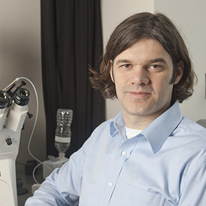
Dr. Reddien is associate professor and associate department head of biology at the Massachusetts Institute of Technology and member of the Whitehead Institute for Biomedical Research. He was an HHMI early career scientist from 2009 to 2014. Continue Reading

More Talks in Development and Stem Cells

  • Martin Raff Part 1
    Control of Cell Growth in Animal Development
  • Cynthia Kenyon Part 1 Genes- hat Control Aging
    Aging Genes: Genes and Cells that Determine the Lifespan of C. elegans
  • Nicole LeDouarin
    Role of Neural Crest in Vertebrate Development and Evolution
  • Michael Levine
    Transcriptional Precision in Drosophila Embryos: Enhancers and Repressors

Related Resources

Research paper discussed in this talk: Petersen CP, Reddien PW. Polarized notum Activation at Wounds Inhibits Wnt Function to Promote Planarian Head Regeneration. Science. 2011 May 13; 332(6031): 852-855.

Reader Interactions

Leave a Reply Cancel reply

Your email address will not be published. Required fields are marked *

Primary Sidebar

Sign up for the Science Communication Lab education newsletter

  • Sign up Newsletter
  • This field is for validation purposes and should be left unchanged.

Privacy Policy

Help us keep bringing the world’s best biology to you!

Footer

Funders

NSF
NIGMS
Lasker
Rita Allen

Start Here

  • Talks for Everyone
  • Talks for Students
  • Talks for Research
  • Talks for Educators

Explore

  • Explore
  • All Playlists
  • All Speakers
  • All Talks

Talks By Topic

  • Biochemistry
  • Bioengineering
  • Biophysics
  • Cell Biology
  • Development and Stem Cells
  • Ecology
  • Genetics and Gene Regulation
  • Human Disease
  • Immunology
  • Microbiology
  • Neuroscience
  • Plant Biology
  • Techniques

Talks by Series

  • Bench to Bedside
  • Famous Discoveries
  • Great Questions
  • Share Your Research Series

Career

  • Professional Development
  • Career Exploration
  • NRMN Resources
  • Biomedical Workforce

Courses

  • Microscopy Series
  • Short Microscopy Series
  • Open edX Courses
  • Cell Biology Flipped Course
  • Engineering Life Flipped Course
  • Evolution Flipped Course

Educator

  • Educator Registration
  • Educator Resources
  • Log In

About Us

  • About Us
  • iBiology Team
  • Wonder Collaborative
  • Contact Us
  • Mission
  • Privacy Policy
  • SCL Financial Conflict of Interest Policy

This material is based upon work supported by the National Science Foundation and the National Institute of General Medical Sciences under Grant No. 2122350 and 1 R25 GM139147. Any opinion, finding, conclusion, or recommendation expressed in these videos are solely those of the speakers and do not necessarily represent the views of the Science Communication Lab/iBiology, the National Science Foundation, the National Institutes of Health, or other Science Communication Lab funders.

© 2023 - 2006 iBiology · All content under CC BY-NC-ND 3.0 license · Privacy Policy · Terms of Use · Usage Policy
 

Power by iBiology