• Skip to primary navigation
  • Skip to main content
  • Skip to primary sidebar
  • Skip to footer

iBiology

Bringing the World's Best Biology to You

  • Start Here
    • Explore iBiology
    • All Playlists
    • All Speakers
    • All Talks
    • What’s new at iBiology?
  • Research Talks
    • Talks by Topic
      • Biochemistry
      • Bioengineering
      • Biophysics
      • Cell Biology
      • Development and Stem Cells
      • Ecology
      • Evolution
      • Genetics and Gene Regulation
      • Human Disease
      • Immunology
      • Microbiology
      • Neuroscience
      • Plant Biology
      • Techniques
      • Archive
    • Talks by Series
      • Bench to Bedside
      • Famous Discoveries
      • Great Unanswered Questions
      • Microscopy Series
      • Share Your Research Series
  • Stories
    • Background to Breakthrough
    • Interviews and Profiles
    • Science and Society
  • Courses
  • Careers
    • Professional Development Talks
    • Professional Development Courses
    • Career Exploration
    • NRMN Resources
    • Biomedical Workforce
  • Educators
  • About
    • Mission
    • Commitment to Inclusion
    • iBiology Team
    • Board of Directors
    • iBiology Funders and Partnerships
    • Contact Us
  • Donate
Home » Courses » Microscopy Series » Image Formation

Correcting for Spherical Aberration with a Correction Collar

  • Duration: 5:22
  • Downloads
    • Hi-Res
    • Low-Res
  • Subtitles
    • English
  • Transcript

IN TRANSCRIPT
00:00:11;26 In this tip, we’re gonna talk a little bit more
00:00:14;02 about spherical aberration,
00:00:15;14 and we’re structural talk specifically about
00:00:18;01 how to minimize or eliminate spherical aberration
00:00:19;22 in the laboratory under the imaging conditions
00:00:22;04 that you’re working on.
00:00:23;22 Just to review, spherical aberration,
00:00:25;08 as shown in this figure here,
00:00:27;15 is the uneven focus of light, axially,
00:00:30;21 from light that passes through the periphery of the lens
00:00:33;10 — that focuses closer to the lens —
00:00:35;06 or light that passes through the axis of the lens —
00:00:38;27 focusing further away from the lens.
00:00:40;25 Spherical aberration,
00:00:43;13 as I had mentioned in the previous lecture,
00:00:44;23 is one of those aberrations that’s extremely difficult,
00:00:48;12 in fact impossible, to completely correct for
00:00:51;14 in the manufacture of lenses
00:00:52;29 because the design criteria that we have to use
00:00:56;19 to make lenses don’t necessarily match the imaging conditions
00:01:00;04 that you need to use in the laboratory
00:01:02;06 to address the questions of interest.
00:01:04;07 Okay?
00:01:05;21 So, we have this paraxial focus
00:01:08;02 and peripheral focus,
00:01:09;26 and we need to bring those together.
00:01:12;03 So, there’s a number of ways to address that, okay?
00:01:17;10 So, one can be utilizing different refractive index immersion media.
00:01:22;04 So, basically, what you’re doing to correct for spherical aberration,
00:01:25;28 no matter how you do it,
00:01:27;27 is adjusting the optical path length
00:01:32;05 to best fit the design criteria of the lens.
00:01:36;17 And we can do that with immersion media,
00:01:38;23 and there are actually kits that you can buy
00:01:40;11 which have many, many different refractive index immersion media
00:01:44;05 that vary at the third decimal place —
00:01:47;22 say, 1.515, 1.516, 1.517, and so on.
00:01:51;28 And you can look in the specimen
00:01:54;09 at the depth that you need to address a question
00:01:58;06 — say you’re looking at the endoplasmic reticulum,
00:02:01;03 and you want to then focus to what looks like
00:02:05;12 it’s something that’s at the limit of resolution
00:02:07;06 close to that depth in the cell —
00:02:09;08 and then test different immersion media
00:02:11;22 at that focus depth
00:02:14;03 so that you get the best correction for spherical aberration.
00:02:17;04 And again, the best correction is found when you see,
00:02:20;11 focusing through, up and down,
00:02:22;18 you have the best symmetry of in and out of focus light, okay?
00:02:25;27 So, you can use this immersion media
00:02:28;10 to correct for spherical aberration
00:02:30;23 and oftentimes that works pretty well.
00:02:36;05 It’s a bit tedious and can be a little messy as well.
00:02:39;28 Another thing you can do is to go ahead
00:02:47;08 and measure your coverslips.
00:02:49;00 To get the design criteria of 170 microns thickness of coverslip,
00:02:54;29 you’ll use a micrometer to measure them
00:02:58;21 and then throw out ones that are too far outside the criteria
00:03:01;22 that you need to have the proper optical path length.
00:03:04;12 In addition to that, you’ll also want to go ahead
00:03:08;03 and, ideally, use a lens with a correction collar,
00:03:11;24 which is really the easiest way to correct for spherical aberration.
00:03:14;27 There are limits, though.
00:03:16;13 So, you have to understand
00:03:18;10 if you’re outside the range that you can correct for,
00:03:20;20 you’ll have to go to these other methods.
00:03:22;10 But a correction collar,
00:03:24;07 which was traditionally available for adjustment of coverslip thickness
00:03:28;25 in air and water immersion lenses,
00:03:30;27 is now also available in oil immersion lenses.
00:03:35;04 And that’s because oil
00:03:37;02 that you use for oil immersion lenses
00:03:38;23 actually has a temperature dependence
00:03:42;07 to its refractive index —
00:03:43;26 as it warms up, the refractive index decreases.
00:03:47;16 So, a correction collar
00:03:50;09 actually works by changing the angle
00:03:54;06 of the peripheral rays versus the axial rays
00:03:57;07 in the objective lens.
00:03:58;25 And you can see the objective lens cut away here.
00:04:00;25 The way a correction collar works
00:04:02;17 is we take the area
00:04:05;23 where you have the steepest angle,
00:04:07;15 right here in the front,
00:04:10;04 between two lens groups in the ray trace,
00:04:12;04 and we take all of the lenses behind that angle
00:04:15;10 — say, here back —
00:04:17;25 we put them in a brass tube,
00:04:19;22 we score a groove around that brass tube,
00:04:22;02 put a pin in it, attach a collar to that,
00:04:25;02 and then we fix the other lenses in another brass tube.
00:04:28;06 When we turn that collar, it moves all of those rear lenses
00:04:32;02 back and forth relative to the front lenses,
00:04:34;22 and those steep angles there then go like this,
00:04:38;13 so that you’re adjusting the peripheral rays
00:04:40;24 while the axial rays don’t get changed quite as much.
00:04:45;29 And that’s exactly how a correction collar works.
00:04:49;10 And then it will bring the paraxial focus,
00:04:51;14 which was further away,
00:04:53;16 back to the peripheral focus, shown here.
00:04:55;22 Okay? So, that’s basically spherical aberration.

This Talk
Speaker: Steve Ross
Audience:
  • Researcher
Recorded: March 2012
More Talks in Microscopy Series
  • Darkfield and Phase Contrast Microscopy (Edward Salmon)
    Darkfield and Phase Contrast Microscopy
  • Shinya Inoue Polarized Light and its Interaction with Material
    Polarized Light and its Interaction with Material
  • Polarization (Edward Salmon)
    Polarization Microscopy
All Talks in Microscopy Series
Share

Talk Overview

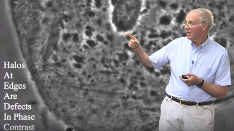
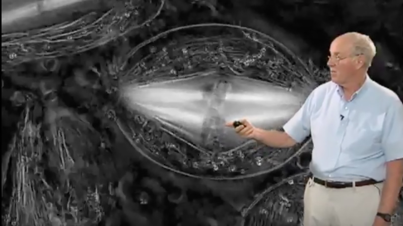
Many microscope objectives are equipped with collars that correct for spherical aberration induced by varying optical thicknesses of the sample substrate, distance between the sample and the substrate or temperature changes. This video tip describes how you set this correction collar to the position optimal for your sample.

Speaker Bio

Steve Ross

Steve Ross

Stephen Ross is the General Manager of Product and Marketing at Nikon Instruments.  He is also very involved in teaching microscopy at the Marine Biological Laboratory in Woods Hole and at the Bangalore Microscopy Course at the National Centre for Biological Sciences. Continue Reading

Playlist: Microscopy Series

  • Adjusting the Eyepiece and Camera Nico Stuurman
    How to Adjust the Eyepiece and Camera
  • Darkfield and Phase Contrast Microscopy (Edward Salmon)
    Darkfield and Phase Contrast Microscopy
  • Shinya Inoue Polarized Light and its Interaction with Material
    Polarized Light and its Interaction with Material
  • Polarization (Edward Salmon)
    Polarization Microscopy

Reader Interactions

Leave a Reply Cancel reply

Your email address will not be published. Required fields are marked *

Primary Sidebar

Sign up for the Science Communication Lab education newsletter

  • Sign up Newsletter
  • This field is for validation purposes and should be left unchanged.

Privacy Policy

Help us keep bringing the world’s best biology to you!

Footer

Funders

NSF
NIGMS
Lasker
Rita Allen

Start Here

  • Talks for Everyone
  • Talks for Students
  • Talks for Research
  • Talks for Educators

Explore

  • Explore
  • All Playlists
  • All Speakers
  • All Talks

Talks By Topic

  • Biochemistry
  • Bioengineering
  • Biophysics
  • Cell Biology
  • Development and Stem Cells
  • Ecology
  • Genetics and Gene Regulation
  • Human Disease
  • Immunology
  • Microbiology
  • Neuroscience
  • Plant Biology
  • Techniques

Talks by Series

  • Bench to Bedside
  • Famous Discoveries
  • Great Questions
  • Share Your Research Series

Career

  • Professional Development
  • Career Exploration
  • NRMN Resources
  • Biomedical Workforce

Courses

  • Microscopy Series
  • Short Microscopy Series
  • Open edX Courses
  • Cell Biology Flipped Course
  • Engineering Life Flipped Course
  • Evolution Flipped Course

Educator

  • Educator Registration
  • Educator Resources
  • Log In

About Us

  • About Us
  • iBiology Team
  • Wonder Collaborative
  • Contact Us
  • Mission
  • Privacy Policy
  • SCL Financial Conflict of Interest Policy

This material is based upon work supported by the National Science Foundation and the National Institute of General Medical Sciences under Grant No. 2122350 and 1 R25 GM139147. Any opinion, finding, conclusion, or recommendation expressed in these videos are solely those of the speakers and do not necessarily represent the views of the Science Communication Lab/iBiology, the National Science Foundation, the National Institutes of Health, or other Science Communication Lab funders.

© 2023 - 2006 iBiology · All content under CC BY-NC-ND 3.0 license · Privacy Policy · Terms of Use · Usage Policy
 

Power by iBiology