Talk Overview
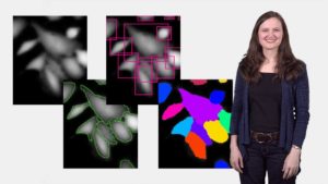
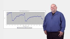
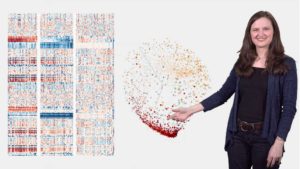
In this series, Dr. Anne Carpenter and Dr. Kevin Eliceiri provide an overview of bioimage analysis. Pre-processing is the first step that follows image acquisition and will prepare your image by reducing the signal-to-noise ratio, applying appropriate filters to the image, and color extraction. Once you perform pre-processing, you’re ready for segmentation, the process of identifying individual cells or structures within an image. If appropriate for your dataset, you can use tracking to be able to link objects in space and time and measure speed, directionality, and cell division. The last step of bioimage analysis is to analyze the data by measuring different features like the number of cells or biological structures, or their size, shape, intensity or texture. Carpenter and Eliceiri finalize this series by providing tips on best practices that will aid scientists in properly analyzing their data.
Concepts:
pre-processing (cropping, color inversion, filtering, color extraction, z-projections, illumination correction, signal to noise, image registration, stitching, deconvolution), segmentation (thresholding, seed-based watershedding, untangling, supervised machine learning, deep learning), tracking, and measurement and phenotype classification (intensity, texture (smoothness), spatial relationships)
Questions
Pre-processing:
- Name three pre-processing techniques and describe what each does to an image.
- Why do striped horizontal and vertical lines often appear in images that are stitched? What can be done to prevent them?
- What is occuring when carrying out Deconvolution? And what is the role of the PSF?
Segmentation:
- Define segmentation and name a few biological situations where you might need/want to segment an image.
- Once you have objects segmented, what are some methods for manipulating those found objects?
- What type(s) of imaging modalities’ images might be better assessed using machine learning as opposed to standard thresholding and watershed techniques?
- What are the pros and cons for using machine learning methods for segmentation?
Tracking:
- What are the three main steps in a typical Tracking analysis?
- Name at least 2 biological situations where you might need/want to track cells.
Measurement:
- Name 3 categories of morphological metrics that can be extracted from biological images.
- True or false? You don’t need to worry about illumination artifacts or bleaching effects during image acquisition because they can be corrected using image analysis methods.
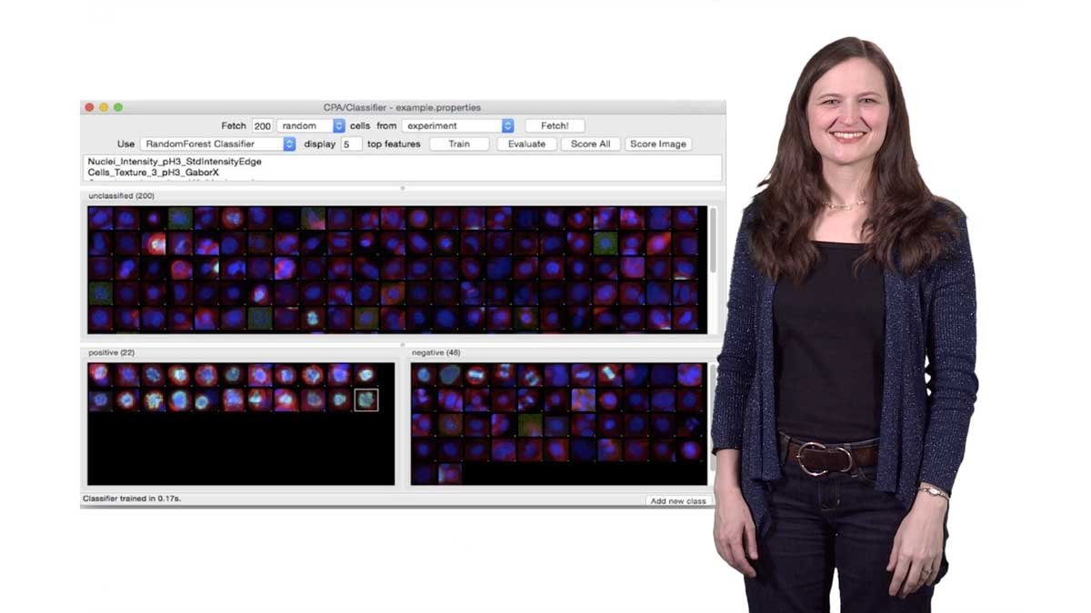
- What is a ‘classifier’ used in supervised machine learning for classifying phenotypes in images. How is it generated?
Best Practices:
- Name the three key “Best Practices” mentioned by Kevin and Anne. Also describe their importance.
Answers
View AnswersSpeaker Bio
Kevin Eliceiri
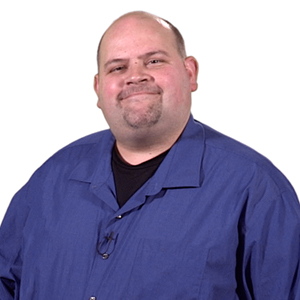
Dr. Kevin Eliceiri is an Associate Professor of Medical Physics and Biomedical Engineering and director of the Laboratory for Optical and Computational Instrumentation (LOCI) at the University of Wisconsin-Madison. Eliceiri completed his bachelor’s and doctoral degree in Biotechnology and Biomedical Engineering at the University of Wisconsin in Madison. In 2008, he founded LOCI and started… Continue Reading
Anne Carpenter
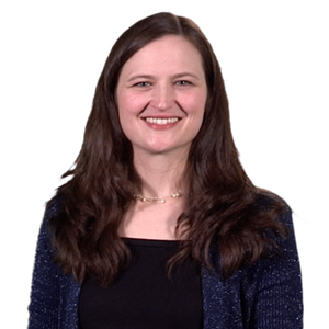
Anne Carpenter is an Institute Scientist at the Broad Institute of MIT and Harvard. Carpenter completed her bachelor’s degree in biological sciences, and a doctoral degree in cell biology from the University of Illinois, Urbana-Champaign. She continued her scientific training as a postdoctoral fellow at the MIT/Whitehead Institute for Biomedical Research in the laboratory of… Continue Reading


Leave a Reply