• Skip to primary navigation
  • Skip to main content
  • Skip to primary sidebar
  • Skip to footer

iBiology

Bringing the World's Best Biology to You

  • Start Here
    • Explore iBiology
    • All Playlists
    • All Speakers
    • All Talks
    • What’s new at iBiology?
  • Research Talks
    • Talks by Topic
      • Biochemistry
      • Bioengineering
      • Biophysics
      • Cell Biology
      • Development and Stem Cells
      • Ecology
      • Evolution
      • Genetics and Gene Regulation
      • Human Disease
      • Immunology
      • Microbiology
      • Neuroscience
      • Plant Biology
      • Techniques
      • Archive
    • Talks by Series
      • Bench to Bedside
      • Famous Discoveries
      • Great Unanswered Questions
      • Young Scientist Series
  • Stories
    • Background to Breakthrough
    • Interviews and Profiles
    • Science and Society
  • Courses
  • Careers
    • Professional Development Talks
    • Professional Development Courses
    • Career Exploration
    • NRMN Resources
    • Biomedical Workforce
  • Educator
  • About
    • Mission
    • iBiology Team
    • Board of Directors
    • Make a Donation
    • iBiology Funders and Partnerships
    • Contact Us
Home » Courses » Microscopy Series » Image Formation

Cleaning a Microscope: How to Find Dirt in Your Optical System

  • Duration: 6:01
  • Downloads
    • Hi-Res
    • Low-Res
  • Subtitles
    • English
  • Transcript

00:00:11.21 Hello, my name is Steve Ross,
00:00:14.03 and today what I want to do is
00:00:17.04 go over some simple troubleshooting
00:00:18.17 with the microscope. And basically talk about
00:00:21.21 how to take care of one of things that's most
00:00:25.21 destructive, but simplest to fix in a microscope system,
00:00:29.02 and that's how to find dirt in the optical system.
00:00:31.10 Like many of the longer tutorials
00:00:37.06 a lot of it has to do with taking advantage
00:00:39.26 of your knowledge of the image planes of the microscope.
00:00:42.14 So you can see right here, I have a microscope image
00:00:44.26 And this is a line up of diatoms
00:00:48.29 on a microscope slide, which are very nice,
00:00:52.01 because they are transparent and you can see
00:00:54.26 them by contrast and there is -- should be a nice,
00:00:59.13 clean, white background on this slide.
00:01:01.23 But you can see that there's an awful lot of dirt
00:01:04.00 in this image. So, one of the first things you do
00:01:07.25 in any type of imaging is to make sure
00:01:10.14 in transmitted light, that the microscope is
00:01:14.00 in proper Kohler alignment.
00:01:15.22 And what that's going to do
00:01:19.00 if you're not in proper alignment,
00:01:20.16 the dirt that's in the aperture planes
00:01:22.07 of the microscope, that shouldn't normally be visible
00:01:24.20 in the image planes, will be visible.
00:01:26.27 So let's see if we're in Kohler alignment.
00:01:28.18 We'll do that by closing down our field aperture,
00:01:31.26 generally what we want to do is make sure that
00:01:34.29 the field aperture is centered.
00:01:38.15 And in focus, meaning that our condenser
00:01:43.02 is in proper focus. So let's adjust this condenser
00:01:46.05 here, with that close field aperture.
00:01:49.10 We can see it was not in focus.
00:01:51.16 Come in closer, closer. And there we have sharp
00:01:55.17 focus of the field of aperture
00:01:57.29 Looks reasonably centered, so let's just
00:01:59.22 open that up right outside our field of view.
00:02:02.08 And adjust our light, there we go.
00:02:06.07 So you can see that did eliminate a good bit of dirt.
00:02:08.18 that was in the optical system.
00:02:10.10 But not all of it, so now you can still see the
00:02:14.11 diatoms. Touch up our fine focus here a little bit.
00:02:18.09 And the second thing we'll do is check whether
00:02:24.05 the dirt is on our camera. Okay, just making sure it's still in sharp focus.
00:02:29.12 Whether it's on our camera or
00:02:33.00 on our specimen, because those are the two image
00:02:35.05 planes that are really greatly affected.
00:02:38.00 So the way we check to see if the dirt is on the camera
00:02:41.05 is by actually rotating the camera.
00:02:43.15 So let's loosen up the camera in the C-mount
00:02:46.25 and rotate that, and what you can see
00:02:50.03 is some dirt in the specimen is rotating
00:02:54.15 relative to the camera when I turn the camera.
00:02:56.13 But that there's a lot of dirt
00:02:58.22 that is actually not moving, and that
00:03:00.28 means it's actually on the window
00:03:03.22 or the camera detector.
00:03:05.14 So let's take the camera off the microscope now.
00:03:11.08 Just loosen that up a bit.
00:03:14.13 And lift this camera out.
00:03:17.12 Remove our iso C-mount here.
00:03:24.09 And I'll take an air duster
00:03:32.20 and blow away the dirt that's on the chip of the camera.
00:03:43.17 Okay, so that should've made a big difference.
00:03:47.14 Let's put that back on the microscope.
00:03:52.02 Okay. And now let's take a look.
00:04:03.24 And rotating the camera now.
00:04:06.15 You see that most of the dirt that's left is rotating with the camera.
00:04:10.12 There's some very light residual dirt
00:04:14.16 that we would actually have to go and clean the window of the camera
00:04:17.13 with some sort of a solvent and lens cleaner,
00:04:20.04 Like lens paper.
00:04:22.21 So let's tighten this up again. And now,
00:04:26.00 we still see that there's some dirt there.
00:04:28.26 So, chances are that's on the slide.
00:04:30.18 And how would we know that?
00:04:31.19 Well if we move the chamber and the slide
00:04:34.28 you could see that the dirt actually moves with the slide.
00:04:38.06 And focus up, because generally if it's on the coverslip
00:04:41.28 it's going to be above the specimen,
00:04:44.06 and sure enough, there's all that dirt that's on our specimen.
00:04:47.06 So let's now take our dust blower
00:04:52.23 and blow out the dirt that's on that back.
00:05:00.24 Okay. that should have removed the majority of the dirt there.
00:05:11.28 And now when we go back in focus
00:05:15.18 we can see that we really eliminated most of the dirt.
00:05:19.14 Again, you're would want to use something
00:05:22.17 that's not going to damage the specimen,
00:05:24.02 like a piece of lens tissue and some solvent
00:05:26.23 to clean even further.
00:05:28.03 But that gives you a pretty good idea
00:05:29.21 of how you would go about methodically troubleshooting dirt
00:05:33.07 in your optical system. Thank you very much.

This Talk
Speaker: Steve Ross
Audience:
  • Researcher
Recorded: January 2013
More Talks in Microscopy Series
  • Light Sources Nico Stuurman
    Light Sources for Microscopy
  • Adjusting the Eyepiece and Camera Nico Stuurman
    How to Adjust the Eyepiece and Camera
  • Correcting for Spherical Aberration with a Correction Collar Stephen Ross
    Correcting for Spherical Aberration with a Correction Collar
All Talks in Microscopy Series
Share

Talk Overview

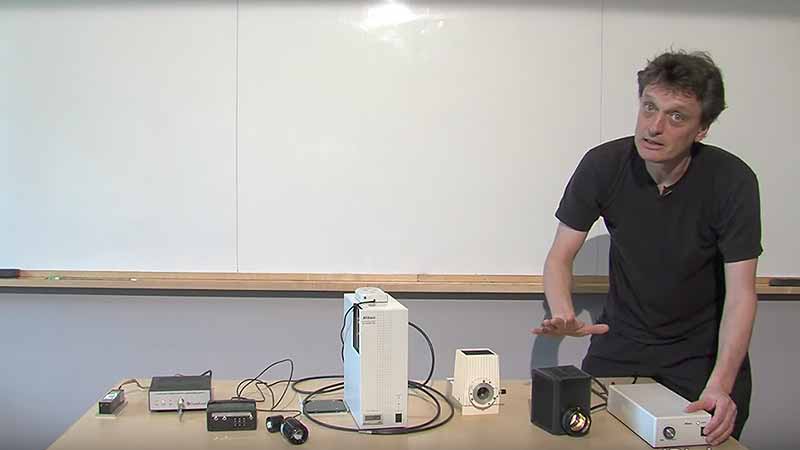
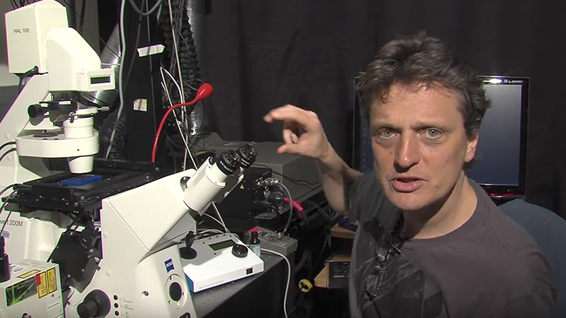
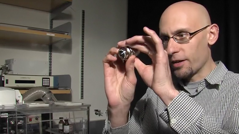
Cleaning a Microscope: Dirt in the microscope degrades image quality. This video explains how an understanding of the optical path helps you find where the dirt is located.

Speaker Bio

Steve Ross

Steve Ross

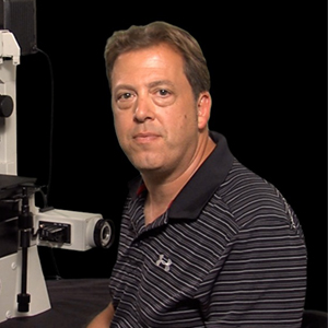
Stephen Ross is the General Manager of Product and Marketing at Nikon Instruments.  He is also very involved in teaching microscopy at the Marine Biological Laboratory in Woods Hole and at the Bangalore Microscopy Course at the National Centre for Biological Sciences. Continue Reading

Playlist: Microscopy Series

  • How To Clean an Objective Lens (Kurt Thorn)
    How to Clean Objective Lenses and Filters
  • Light Sources Nico Stuurman
    Light Sources for Microscopy
  • Adjusting the Eyepiece and Camera Nico Stuurman
    How to Adjust the Eyepiece and Camera
  • Correcting for Spherical Aberration with a Correction Collar Stephen Ross
    Correcting for Spherical Aberration with a Correction Collar

Reader Interactions

Leave a Reply Cancel reply

Your email address will not be published. Required fields are marked *

Primary Sidebar

Like our Website?

You'll love our newsletter...

  • Sign up for:
    • Exclusive iBiology content
    • 10 Must See Biology Videos
    • Talks by 20+ Nobel Winners
    • Our young Scientist Survival Toolkit
    • Talks by trailblazing Women in Biology
    • Updates on exciting iBiology projects in development
    Tyler Allen
  • This field is for validation purposes and should be left unchanged.

Privacy Policy

Help us keep bringing the world’s best biology to you!

Footer

Funders

NSF Logo
Lasker Logo
NIGMS Logo

Partners

Start Here

  • Talks for Everyone
  • Talks for Students
  • Talks for Research
  • Talks for Educators

Explore

  • Explore
  • All Playlists
  • All Speakers
  • All Talks

Talks By Topic

  • Biochemistry
  • Bioengineering
  • Biophysics
  • Cell Biology
  • Development and Stem Cells
  • Ecology
  • Genetics and Gene Regulation
  • Human Disease
  • Immunology
  • Microbiology
  • Neuroscience
  • Plant Biology
  • Techniques

Talks by Series

  • Bench to Bedside
  • Famous Discoveries
  • Great Questions
  • Share Your Research Series

Career

  • Professional Development
  • Career Exploration
  • NRMN Resources
  • Biomedical Workforce

Courses

  • Microscopy Series
  • Short Microscopy Series
  • Open edX Courses
  • Cell Biology Flipped Course
  • Engineering Life Flipped Course
  • Evolution Flipped Course

Educator

  • Educator Registration
  • Educator Resources
  • Log In

About Us

  • About Us
  • iBiology Team
  • Wonder Collaborative
  • Contact Us
  • Make a Donation
  • Mission
  • Privacy Policy

This material is based upon work supported by the National Science Foundation and the National Institute of General Medical Sciences under Grant No. MCB-1052331. Any opinion, finding, conclusion, or recommendation expressed in these videos are solely those of the speaker and do not necessarily represent the views of iBiology, the National Science Foundation, the National Institutes of Health, or other iBiology funders.

© 2021 - 2006 iBiology · All content under CC BY-NC-ND 3.0 license · Privacy Policy · Terms of Use · Usage Policy
 

Power by iBiology


This website uses cookies to remember you and improve your experience. To find out more see our Privacy Policy.Accept