• Skip to primary navigation
  • Skip to main content
  • Skip to primary sidebar
  • Skip to footer

iBiology

Bringing the World's Best Biology to You

  • Start Here
    • Explore iBiology
    • All Playlists
    • All Speakers
    • All Talks
    • What’s new at iBiology?
  • Research Talks
    • Talks by Topic
      • Biochemistry
      • Bioengineering
      • Biophysics
      • Cell Biology
      • Development and Stem Cells
      • Ecology
      • Evolution
      • Genetics and Gene Regulation
      • Human Disease
      • Immunology
      • Microbiology
      • Neuroscience
      • Plant Biology
      • Techniques
      • Archive
    • Talks by Series
      • Bench to Bedside
      • Famous Discoveries
      • Great Unanswered Questions
      • Microscopy Series
      • Share Your Research Series
  • Stories
    • Background to Breakthrough
    • Interviews and Profiles
    • Science and Society
  • Courses
  • Careers
    • Professional Development Talks
    • Professional Development Courses
    • Career Exploration
    • NRMN Resources
    • Biomedical Workforce
  • Educators
  • About
    • Mission
    • iBiology Team
    • Board of Directors
    • iBiology Funders and Partnerships
    • Contact Us
  • Donate
Home » Courses » Microscopy Series » Image Formation

Light Sources for Microscopy

  • Duration: 7:19
  • Downloads
    • Hi-Res
    • Low-Res
  • Subtitles
    • English
  • Transcript

00:00:11.23 I'd like to show you some of the light sources that we are using
00:00:16.20 nowadays in microscopy. And the very first light source that we don't use
00:00:21.12 anymore on modern microscopes is right outside. The sun has been
00:00:26.18 used for many centuries to illuminate our specimens, often using
00:00:32.08 a little mirror that then directs the sunlight onto the specimen.
00:00:35.21 So that used to be the brightest light source we could find.
00:00:39.03 Nowadays, we're more inside and we're using devices like those standing here.
00:00:45.11 So first of all, here on the left, I have a halogen lamp. The lamp
00:00:53.15 itself is sitting in a lamp house. It is a halogen lamp, very much like
00:00:59.07 what you use at home. We can put a voltage on this, we'll then
00:01:05.07 illuminate -- the spiral here will start glowing, and that light will
00:01:11.12 then through a condenser lens be transmitted into our microscope.
00:01:16.03 So some of the halogen lamp houses have a little mirror sitting here on the back
00:01:21.24 to get more light coming out of the front. Often that's not needed,
00:01:26.00 this lamp house here is just completely black inside and all you get out is the
00:01:33.12 light coming out of the front. Since we have a condenser lens here, in front
00:01:38.05 of the filaments, that lens will make a magnified image of the filament.
00:01:44.00 And I project it here on the whiteboard, and so that image of the filament,
00:01:49.11 it's the image of our light source, is something you will also encounter in
00:01:54.16 various positions throughout the microscope. Now halogen lamps
00:01:59.17 are nice for transmitted light illumination, but they have two disadvantages. One is
00:02:05.08 that as you see, it takes quite a bit of time for the light to come on. It doesn't
00:02:10.29 switch on momentarily, and the other thing is it doesn't get super
00:02:15.04 bright. And for things like fluorescence, we really want brighter light sources.
00:02:20.05 So, a light source that has been used for many, many years in fluorescence microscopy
00:02:27.16 is this mercury arc lamp. This is the bulb, and in this bulb, there is an anode and a
00:02:37.14 cathode, and a very high voltage is being put over this such that it forms
00:02:43.13 an arc. Just the same thing as when you're welding, which of course everyone
00:02:48.23 does in their spare time. And this arc is super, super bright. That arc is
00:02:56.13 then projected so that the light goes in every direction through a condenser
00:03:02.01 lens it reaches our microscope. Also, inside this lamp house, there's often a mirror,
00:03:08.03 a parabolic mirror and that reflects the light going into the back direction
00:03:13.04 so that it comes out in the front. So you get more light coming out.
00:03:16.12 So this is a mercury arc lamp. We don't like them that much anymore
00:03:24.01 and that is because you have to replace them after about 200 hours.
00:03:27.18 Also when these things break, they're a big hazard. Immediately
00:03:32.13 evacuate the room if that happens. And they're kind of a pain to
00:03:36.29 align. You have to set them up so that this arc is positioned in a right
00:03:41.20 place, with respect to the condenser lens. So that the light goes
00:03:46.00 exactly through the optical path of the microscope. Therefore, little
00:03:51.08 boxes like this one here have become more and more popular. Here,
00:03:56.09 we have a so-called metal halide illuminator, it's also a lamp.
00:04:04.02 And that thing just clicks in here, there's no alignment that needs to be done.
00:04:09.12 The light is then delivered through this thing here, which is a liquid
00:04:15.06 light guide. So it's a tube that's filled with a liquid, something like water,
00:04:22.08 and the light is coupled in on this end and makes it all the way to the
00:04:27.28 end. So when I now assemble this and fire it up here.
00:04:38.14 Makes a lot of noise, takes some time. So one thing about these
00:04:44.26 lamps is that they don't like to be switched on and off very often.
00:04:49.14 So you know, once they're on, leave them on for at least an hour or so.
00:04:53.29 So when I now press the shutter, so that it opens, you see the light
00:04:58.16 coming out of this liquid light guide. The lamp needs to warm up a bit,
00:05:03.21 and it will become brighter and brighter over time. The light source
00:05:07.15 that is more and more replacing both the halogens and things like this
00:05:11.28 is an LED. And here I have one of those light emitting diodes. So
00:05:17.25 these things are really nice because you switch them on very, very rapidly,
00:05:23.03 there's no fans, much less heat production. And nowadays, they can
00:05:29.10 become very, very bright, as well. So often when you couple this into
00:05:35.10 the microscope, you will put a condenser lens in front of it. And you will get an assembly
00:05:40.03 something like this. And we have nice light of the wavelength that we're
00:05:44.20 interested in coming out of them. So these LEDS have defined colors, but
00:05:50.28 you can get multiples of them and combine them, and electronically
00:05:55.13 select which wavelength you would like to use.
00:05:59.14 Lastly, in quite a few microscopies, such a confocal and TIRF,
00:06:06.25 we use lasers. Lasers can look like this one here. So this is a
00:06:14.00 solid state laser. So as you know, a laser is really a narrow beam
00:06:19.07 of light coming out. And I'll switch this one on. So again, this takes
00:06:24.23 a little bit of time to warm up. Lasers, since they produce this very
00:06:29.28 narrow beam of light, you have to really watch out with them. You
00:06:34.16 don't want to get that beam of light hitting your eyeball ever.
00:06:38.27 This has a safety shutter here in the front, and so you can now see
00:06:43.25 this very bright spot of green laser light coming out.
00:06:48.06 Okay, that was the various types of light sources that we use
00:06:53.12 with microscopes.

This Talk
Speaker: Nico Stuurman
Audience:
  • Researcher
Recorded: April 2012
More Talks in Microscopy Series
  • Adjusting the Eyepiece and Camera Nico Stuurman
    How to Adjust the Eyepiece and Camera
  • Correcting for Spherical Aberration with a Correction Collar Stephen Ross
    Correcting for Spherical Aberration with a Correction Collar
  • Darkfield and Phase Contrast Microscopy (Edward Salmon)
    Darkfield and Phase Contrast Microscopy
All Talks in Microscopy Series
Share

Talk Overview

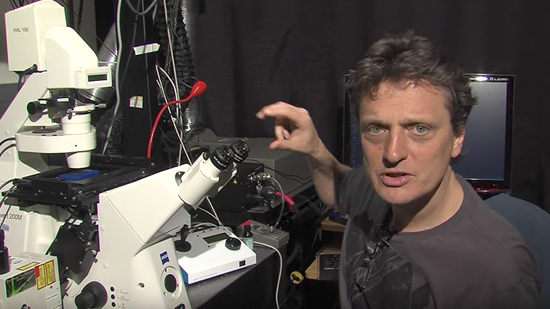
Most light sources under the sun (sun included) can be used in microscopy. In this talk about light sources for microscopy, Nico Stuurman shows a few examples of light sources that are currently used in microscopes, including Halogen lamps, Metal Halide burners, LEDs, and lasers.

Speaker Bio

Nico Stuurman

Nico Stuurman

Nico Stuurman is a Research Specialist at the University of California, San Francisco, in the lab of Ron Vale. Nico combines his expertise in computer programming and microscopy to advance many projects including the Open Source software, Micro-Manager. Continue Reading

Playlist: Microscopy Series

  • Stephen Ross
    Cleaning a Microscope: How to Find Dirt in Your Optical System
  • Adjusting the Eyepiece and Camera Nico Stuurman
    How to Adjust the Eyepiece and Camera
  • Correcting for Spherical Aberration with a Correction Collar Stephen Ross
    Correcting for Spherical Aberration with a Correction Collar
  • Darkfield and Phase Contrast Microscopy (Edward Salmon)
    Darkfield and Phase Contrast Microscopy

Reader Interactions

Comments

  1. Linda says

    September 14, 2021 at 5:17 am

    What do you think that Is a light microscope better than a conventional one?

    Reply

Leave a Reply Cancel reply

Your email address will not be published. Required fields are marked *

Primary Sidebar

Like our Website?

You'll love our newsletter...

  • Sign up for:
    • Exclusive iBiology content
    • 10 Must See Biology Videos
    • Talks by 20+ Nobel Winners
    • Our young Scientist Survival Toolkit
    • Talks by trailblazing Women in Biology
    • Updates on exciting iBiology projects in development
    Tyler Allen
  • This field is for validation purposes and should be left unchanged.

Privacy Policy

Help us keep bringing the world’s best biology to you!

Footer

Funders

National Science Foundation: NSF
Lasker Foundation
NIGMS

Partners

EMBL
EMBO
HHMI
NRMN
UCSF

Start Here

  • Talks for Everyone
  • Talks for Students
  • Talks for Research
  • Talks for Educators

Explore

  • Explore
  • All Playlists
  • All Speakers
  • All Talks

Talks By Topic

  • Biochemistry
  • Bioengineering
  • Biophysics
  • Cell Biology
  • Development and Stem Cells
  • Ecology
  • Genetics and Gene Regulation
  • Human Disease
  • Immunology
  • Microbiology
  • Neuroscience
  • Plant Biology
  • Techniques

Talks by Series

  • Bench to Bedside
  • Famous Discoveries
  • Great Questions
  • Share Your Research Series

Career

  • Professional Development
  • Career Exploration
  • NRMN Resources
  • Biomedical Workforce

Courses

  • Microscopy Series
  • Short Microscopy Series
  • Open edX Courses
  • Cell Biology Flipped Course
  • Engineering Life Flipped Course
  • Evolution Flipped Course

Educator

  • Educator Registration
  • Educator Resources
  • Log In

About Us

  • About Us
  • iBiology Team
  • Wonder Collaborative
  • Contact Us
  • Mission
  • Privacy Policy
  • SCL Financial Conflict of Interest Policy

This material is based upon work supported by the National Science Foundation and the National Institute of General Medical Sciences under Grant No. 2122350 and 1 R25 GM139147. Any opinion, finding, conclusion, or recommendation expressed in these videos are solely those of the speakers and do not necessarily represent the views of the Science Communication Lab/iBiology, the National Science Foundation, the National Institutes of Health, or other Science Communication Lab funders.

© 2022 - 2006 iBiology · All content under CC BY-NC-ND 3.0 license · Privacy Policy · Terms of Use · Usage Policy
 

Power by iBiology