• Skip to primary navigation
  • Skip to main content
  • Skip to primary sidebar
  • Skip to footer

iBiology

Bringing the World's Best Biology to You

  • Start Here
    • Explore iBiology
    • All Playlists
    • All Speakers
    • All Talks
    • What’s new at iBiology?
  • Research Talks
    • Talks by Topic
      • Biochemistry
      • Bioengineering
      • Biophysics
      • Cell Biology
      • Development and Stem Cells
      • Ecology
      • Evolution
      • Genetics and Gene Regulation
      • Human Disease
      • Immunology
      • Microbiology
      • Neuroscience
      • Plant Biology
      • Techniques
      • Archive
    • Talks by Series
      • Bench to Bedside
      • Famous Discoveries
      • Great Unanswered Questions
      • Microscopy Series
      • Share Your Research Series
  • Stories
    • Background to Breakthrough
    • Interviews and Profiles
    • Science and Society
  • Courses
  • Careers
    • Professional Development Talks
    • Professional Development Courses
    • Career Exploration
    • NRMN Resources
    • Biomedical Workforce
  • Educators
  • About
    • Mission
    • iBiology Team
    • Board of Directors
    • iBiology Funders and Partnerships
    • Contact Us
  • Donate
Home » Courses » Microscopy Series » Fluorescence Microscopy

Correlating Fluorescence with Electron Microscopy

  • Duration: 8:08
  • Downloads
    • Hi-Res
    • Low-Res
  • Subtitles
    • English
  • Transcript

00:00:11.18 Finally, I want to go in the other direction. And solve two possible
00:00:16.11 problems. One of them was that, as you remember, GFP
00:00:19.28 absolutely needs the presence of oxygen to mature.
00:00:22.29 What about if you're forced to work on an obligate anaerobe?
00:00:26.22 Also, how do we go to yet higher spatial resolution? And
00:00:32.20 there are tricks to do so with optical microscopy, but the fundamental
00:00:36.09 way that most of our high resolution spatial information in cell biology
00:00:40.24 has come about has been through electron microscopy.
00:00:43.21 And up till now, electron microscopy has lacked the genetically
00:00:47.23 encodable tag that does what GFP does. In other words, where
00:00:52.03 you can just put in a gene, fuse it to the protein you care about,
00:00:55.25 and then follow it around by your favorite form of microscopy. It could
00:00:59.22 be great if we could do that in electron microscopy. And here
00:01:03.03 the trick is again, to go to another completely different protein
00:01:07.06 family. This is Arabidopsis phototropin, which binds flavins, in particular
00:01:12.17 flavin mononucleotide. And flavins are also ubiquitous in biochemistry,
00:01:18.06 and essentially all organisms that we care about have them, including
00:01:21.01 the obligate anaerobes. And so, this protein as it came out of the plant
00:01:27.05 doesn't do any fluorescence, it only uses light to trigger phototransduction,
00:01:32.24 but again, via somewhat similar trick -- a somewhat analogous trick
00:01:37.06 to the infrared fluorescence protein, we can frustrate this signal
00:01:41.03 transduction, turn it into a fluorophore, and this fluorophore
00:01:45.14 happens, by the way, to make something called singlet oxygen
00:01:49.09 as a byproduct. It does it actually quite well, and these curves
00:01:55.13 represent the data showing that the fluorescence switches rather like
00:01:59.16 GFP in wavelengths, but admittedly, not terribly bright.
00:02:03.11 And plus, it's singlet oxygen. Singlet oxygen is normally a bad
00:02:07.22 thing for us. And microscopists don't like it, because it's
00:02:12.05 responsible for a lot of the photobleaching. So we try to make
00:02:15.03 as little singlet oxygen as possible. But when you have a protein
00:02:18.02 that makes singlet oxygen well, you can use a special form of histochemistry
00:02:22.22 that's been known for a long time, and singlet oxygen will polymerize
00:02:29.07 a molecule that we supply in dead fixed cells. So remember, electron
00:02:34.04 microscopy has to be done, and has generally only been done on
00:02:38.00 dead fixed cells. So we don't mind after fixation, supplying diaminobenzidine,
00:02:42.22 and this molecule is instantly polymerized very locally wherever
00:02:47.28 the singlet oxygen is made. It's polymerized into a precipitate
00:02:52.00 that then is stainable by osmium, and osmium is the counterstain that we use so much
00:02:57.07 in electron microscopy anyway. So wherever this protein was fused,
00:03:01.11 when we excite it with light in the presence of oxygen gas, and
00:03:05.23 maybe I should explain here, oxygen gas that you and I are breathing
00:03:09.01 is a very unusual molecule, in the sense that it's a triplet.
00:03:14.08 It has unpaired electrons, and when it encounters the excited
00:03:20.04 state of this singlet oxygen generating protein, the regular oxygen
00:03:26.07 gets excited to this singlet oxygen state, which is an excited state of
00:03:30.14 oxygen still diffusible like regular oxygen. Can cross membranes,
00:03:34.23 but it is ravening beast in its chemical reactivity, and it loves to attack methionines,
00:03:42.08 tryptophans, histidines, and so on. But also will attack diaminobenzidine
00:03:47.03 to make this polymer. So here, we've transfected in miniSOG and targeted
00:03:53.00 the mitochondria by fusing it to a piece from cytochrome c, which is
00:03:59.03 a well known mitochondrial protein. And you can see the mitochondria by fluorescence
00:04:04.12 here. And they look like regular mitochondria. In a live cell they look sort of like
00:04:10.03 these little wispy threads. But then the crucial thing is we can
00:04:15.12 fix the cells and turn up the light and bubble pure oxygen to help
00:04:20.20 efficiency and include diaminobenzidine, and wherever there was fluorescence
00:04:24.20 before, we turn it into this black precipitate, and that's not too impressive yet. But
00:04:31.00 that black precipitate can be looked at under electron microscope at higher
00:04:35.24 and higher magnification. So this is a blowup of one of those mitochondria
00:04:40.12 down to the scale where you can see 200nm and we can see
00:04:44.10 all the cristae and the spaces between the inner and outer membrane.
00:04:48.02 This is the sort of classic appearance of a mitochondrion that you would've seen
00:04:52.09 from a textbook. But this mitochondrion has been picked out genetically.
00:04:55.13 And here is another test case, this is the gap junction that I mentioned.
00:05:00.19 And we fused connexin43, one of the major constituents of gap junctions,
00:05:06.17 to this so-called single oxygen generating protein, miniSOG. And by the way,
00:05:11.18 I forgot to say "mini" refers to the fact that this protein is only 106 amino acids,
00:05:16.16 it's less than half the size of GFP. And there are times when it is
00:05:21.26 a better fusion partner just because it's small. And as I said, it doesn't
00:05:25.19 need oxygen to fluoresce, because it uses the flavins that the cells provide.
00:05:31.03 Of course when we want to make the precipitate, we have to provide
00:05:34.16 oxygen, but that's done anyway, after the cell is dead and fixed.
00:05:38.16 So, these are the gap junctional stripes. These are in ordinary fluorescence
00:05:44.15 microscope level, these sort of boundaries between individual
00:05:47.27 cells that are lit up as gap junctions. This is after we have illuminated them
00:05:53.02 in the presence of diaminobenzidine and oxygen, and converted them
00:05:56.18 into black precipitates. And then when we blow it up, we can
00:06:00.25 blow it up to the scale where we can actually see what we believe individual
00:06:05.23 hexamers. As these white shadows in what looks like the machine gun
00:06:11.13 belt of precipitate. That's its sort of crude appearance. It looks like
00:06:17.07 bullets periodically spaced. And these white blobs may be
00:06:23.13 the leftover connexin that is blocking the formation of precipitate
00:06:26.27 everywhere else. In other words, the miniSOG up here are spitting out
00:06:31.01 precipitate and they got every they can, but the protein where the
00:06:35.00 connexin is sort of blocks it and keeps it away, because the space
00:06:39.11 is already occupied. And then when the knife that makes the section
00:06:43.02 cuts through this, if you happen to cut right through the center
00:06:45.28 of this region, you can get the periodic array and see things at very
00:06:51.01 much higher resolution. By comparison, old fashion techniques, still
00:06:57.02 useful in many cases but more difficult, which is immunogold
00:07:02.00 electron microscopy. Only light captures a very small fraction of the proteins because
00:07:09.00 here, we are trying to diffuse antibodies through a fixed tissue.
00:07:13.22 And the fixation tends to destroy a lot of antigenic reactivity, and
00:07:18.25 antibody has a hard time getting into the section. Also any excess
00:07:23.14 has to be washed out. So we're lucky when we can at least see a few
00:07:26.13 dots at the gap junction. But a picture like this, which is a good quality
00:07:30.20 immuno EM, doesn't give you any impression of how densely
00:07:34.28 packed this crystalline array of connexins is. And we know this
00:07:38.02 from many other experiments, that the connexins look something like this,
00:07:42.04 according to this model that I'm showing up here.

This Talk
Speaker: Roger Tsien
Audience:
  • Educators of Adv. Undergrad / Grad
  • Researcher
  • Educators
Recorded: May 2012
More Talks in Microscopy Series
  • Steven Chu
    Pushing the Boundaries of Light Microscopy
  • Disassembling a Nikon Ti Eclipse Stephen Ross
    Demystifying Microscopes: Disassembling a Nikon Ti Eclipse Microscope
  • Nico Sturman on iBiology: Disassembling and ASI Ramm in Microscopy
    Demystifying Microscopes: Disassembling an ASI RAMM Microscope
All Talks in Microscopy Series
Share

Talk Overview

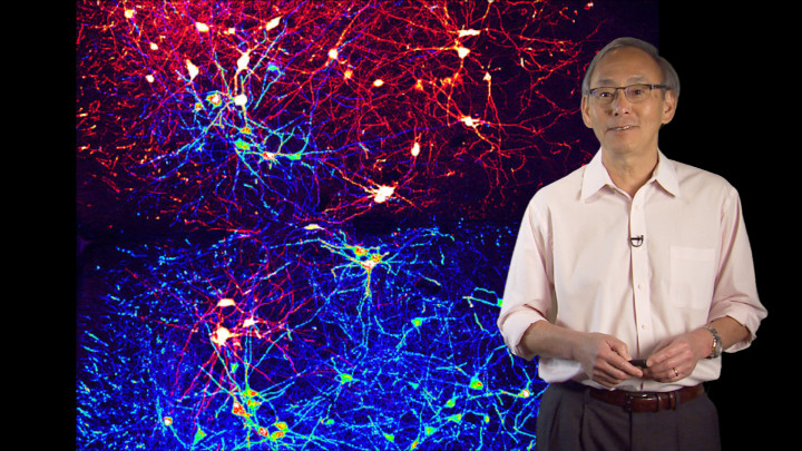
This lecture discusses a new protein, miniSOG, which can be imaged fluorescently but which can also produce an electron-dense deposit for visualization in electron microscopy, thus enabling correlated light and electron microscopy.

Speaker Bio

Roger Tsien

Roger Tsien

Dr. Tsien was a Professor at the University of California, San Diego, a Howard Hughes Medical Institute Investigator and a member of the National Academy of Sciences. In 2008, Tsien shared the Nobel Prize in Chemistry for the discovery and development of green fluorescent protein, GFP. His lab continues to develop new fluorescent proteins as… Continue Reading

Playlist: Microscopy Series

  • Labeling Proteins with Fluorescent Probes (Timothy Mitchison)
    Labeling Proteins with Fluorescent Probes
  • Steven Chu
    Pushing the Boundaries of Light Microscopy
  • Disassembling a Nikon Ti Eclipse Stephen Ross
    Demystifying Microscopes: Disassembling a Nikon Ti Eclipse Microscope
  • Nico Sturman on iBiology: Disassembling and ASI Ramm in Microscopy
    Demystifying Microscopes: Disassembling an ASI RAMM Microscope

Reader Interactions

Leave a Reply Cancel reply

Your email address will not be published. Required fields are marked *

Primary Sidebar

Like our Website?

You'll love our newsletter...

  • Sign up for:
    • Exclusive iBiology content
    • 10 Must See Biology Videos
    • Talks by 20+ Nobel Winners
    • Our young Scientist Survival Toolkit
    • Talks by trailblazing Women in Biology
    • Updates on exciting iBiology projects in development
    Tyler Allen
  • This field is for validation purposes and should be left unchanged.

Privacy Policy

Help us keep bringing the world’s best biology to you!

Footer

Funders

National Science Foundation: NSF
Lasker Foundation
NIGMS

Partners

EMBL
EMBO
HHMI
NRMN
UCSF

Start Here

  • Talks for Everyone
  • Talks for Students
  • Talks for Research
  • Talks for Educators

Explore

  • Explore
  • All Playlists
  • All Speakers
  • All Talks

Talks By Topic

  • Biochemistry
  • Bioengineering
  • Biophysics
  • Cell Biology
  • Development and Stem Cells
  • Ecology
  • Genetics and Gene Regulation
  • Human Disease
  • Immunology
  • Microbiology
  • Neuroscience
  • Plant Biology
  • Techniques

Talks by Series

  • Bench to Bedside
  • Famous Discoveries
  • Great Questions
  • Share Your Research Series

Career

  • Professional Development
  • Career Exploration
  • NRMN Resources
  • Biomedical Workforce

Courses

  • Microscopy Series
  • Short Microscopy Series
  • Open edX Courses
  • Cell Biology Flipped Course
  • Engineering Life Flipped Course
  • Evolution Flipped Course

Educator

  • Educator Registration
  • Educator Resources
  • Log In

About Us

  • About Us
  • iBiology Team
  • Wonder Collaborative
  • Contact Us
  • Mission
  • Privacy Policy
  • SCL Financial Conflict of Interest Policy

This material is based upon work supported by the National Science Foundation and the National Institute of General Medical Sciences under Grant No. 2122350 and 1 R25 GM139147. Any opinion, finding, conclusion, or recommendation expressed in these videos are solely those of the speakers and do not necessarily represent the views of the Science Communication Lab/iBiology, the National Science Foundation, the National Institutes of Health, or other Science Communication Lab funders.

© 2022 - 2006 iBiology · All content under CC BY-NC-ND 3.0 license · Privacy Policy · Terms of Use · Usage Policy
 

Power by iBiology