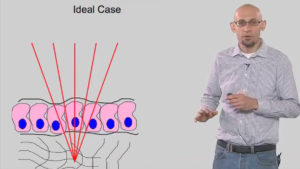
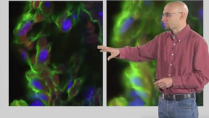
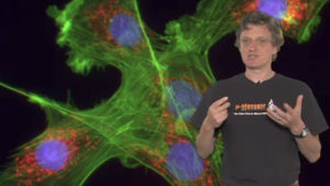
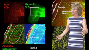
Clare Waterman, developer of fluorescent speckle microscopy, describes computational tools (developed by Gaudenz Danuser) for automatic quantitative analysis of speckle microscopy data. (Talk recorded in July 2012)
Audience:
- Researcher
Duration: 6:16